
X-RAY Right SI Joint Oblique View
Experience the benefits of an X-RAY Right SI Joint Oblique View at Medintu. Our high-quality imaging services, which are at par with the best in the industry, are affordable, ensuring you get the best value for your investment. With our NABL and NABH accreditations, you can trust the precision and reliability of our results, providing you with peace of mind about your health.


Fill out form to enquire now
X-RAY Right SI Joint Oblique View
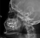
Are you seeking an affordable X-RAY Right SI Joint Oblique View? Medintu offers competitively priced MRI and CT scans, partnering with top NABL-certified diagnostic centers and clinics. Our facilities ensure high-quality imaging and accurate results. An X-ray of the right SI (sacroiliac) joint in an oblique view provides a clear image of the joint’s structure and alignment from a specific angle. This view helps in assessing the sacroiliac joint for any signs of inflammation, injury, or degenerative changes. It is commonly used to diagnose conditions such as arthritis, fractures, or joint dysfunction in the lower back and pelvic region. The oblique angle allows for better visualization of the joint space, helping physicians to detect subtle abnormalities that might not be visible in other views. This imaging technique is valuable for providing a more comprehensive understanding of the patient’s condition and guiding treatment decisions. To book an X-RAY Right SI Joint Oblique View scan appointment, visit our platform, Medintu, or contact us at +919100907036 or +919100907622 for reasonable prices.
Overview
Specification
FAQ
Overview
What is an X-RAY Right SI Joint Oblique View?
An X-ray Right SI Joint Oblique View is a medical imaging technique that captures detailed images of the right sacroiliac (SI) joint from an angled perspective. This view helps healthcare providers evaluate the joint for conditions such as inflammation, fractures, arthritis, or other abnormalities affecting the lower back and pelvis. It is particularly useful for assessing the alignment and structure of the sacroiliac joint and detecting issues that may not be visible from other angles. The oblique angle of the X-ray provides a clearer image of the joint, showing the relationship between the sacrum and iliac bones more effectively. This imaging technique is commonly recommended for individuals experiencing unexplained lower back pain or suspected SI joint dysfunction.
Why is it done?
- To diagnose conditions such as arthritis, fractures, or inflammation in the sacroiliac joint.
- To assess sacroiliac joint dysfunction in patients experiencing lower back or pelvic pain.
- To evaluate any abnormality or injury in the right SI joint, especially after trauma.
- To monitor the progression of diseases like osteoarthritis or ankylosing spondylitis.
- To plan the appropriate treatment for SI joint-related problems, such as physical therapy or surgical interventions.
How do you prepare for an X-ray Right SI Joint Oblique View?
- There are no special preparations needed, but you might be asked to remove clothing or accessories that could interfere with the imaging.
- Wear a hospital gown if instructed to do so by the medical team.
- Inform the technician if you are pregnant or suspect pregnancy, as X-rays are typically avoided during pregnancy unless necessary.
- If the procedure is part of a series of X-rays, follow any specific instructions provided by your doctor.
- Stay calm and follow instructions for positioning and holding still during the procedure.
What is the procedure for an X-ray Right SI Joint Oblique View?
- You will be asked to lie down on your side or stand, depending on the best position for the X-ray.
- The technician will carefully position you to ensure the right SI joint is visible at the oblique angle.
- You may need to adjust your posture or angle slightly to achieve the most accurate view of the joint.
- The technician will take the X-ray image, which may require you to hold still for a few seconds.
What is the aftercare for an X-ray Right SI Joint Oblique View?
- There is typically no aftercare required for this procedure.
- You can return to your usual activities immediately after the X-ray unless otherwise advised by your doctor.
- If you are asked to wear a gown during the procedure, you can change back into your clothes once the X-ray is completed.
- Wait for your doctor to review the images and discuss the results with you.
What is the cost of an X-RAY Right SI Joint Oblique View?
The X-RAY Right SI Joint Oblique View scan costs Rs. 500/- to Rs.1,000/-, and the reports are available within 24 hours, ensuring you receive your results promptly and can take the following steps in your treatment plan.
Specification
- Test Type: X-RAY Right SI Joint Oblique View
Preparation:
Wear a loose-fitting cloth
Fasting is required for a few hours if instructed
Stay hydrated
Carry Your ID Proof
Remove jewellery
As per the PC-PNDT Act, patients must have a prescription with a doctor’s signature, stamp, and DMC/HMC number. This is a crucial requirement that ensures the safety and accuracy of the procedure. Please ensure you have the necessary prescription before booking your appointment.
Reports Time: Within 24 hours
Test Price: starts from Rs. 500/- to Rs.1,000/-
- Test Type: X-RAY Right SI Joint Oblique View
FAQ
- How can I book an appointment for a low-cost X-RAY Right SI Joint Oblique View through Medintu?
Booking an appointment for an affordable X-RAY Right SI Joint Oblique View at Medintu is straightforward. You can do it through our website, Medintu, by filling out the inquiry form or contacting us at +919100907036 or +919100907622. Our team will assist you in finding the earliest available slot that suits your schedule.
- What is an X-RAY Right SI Joint Oblique View Scan?An X-ray Right SI Joint Oblique View is a medical imaging technique that captures detailed images of the right sacroiliac (SI) joint from an angled perspective.
- What is the cost of an X-RAY Right SI Joint Oblique View?
The X-RAY Right SI Joint Oblique View scan costs Rs. 500/- to Rs.1,000/-, and the reports are available within 24 hours.
- When will my reports be available?Reports will be available within 24 hours after your scan.
- Is an X-RAY of the Right SI Joint Oblique View safe?Yes, an X-ray Right SI Joint Oblique View is generally safe, though it involves low levels of radiation, which is carefully controlled.
- What can an X-RAY Right SI Joint Oblique View detect?
- An X-ray Right SI Joint Oblique View can detect conditions such as arthritis, fractures, inflammation, joint dysfunction, and other abnormalities in the sacroiliac joint.
Why Choose Medintu for Your X-RAY Right SI Joint Oblique View?
Medintu is a leading digital healthcare platform that provides high-quality MRI scans for your MRI And SI Joints at affordable prices in trusted laboratories. We collaborate with top-tier labs and clinics with the latest technology to offer a wide range of ultrasound scans with accurate results. Your safety and comfort are our top priorities. We provide various ultrasounds for twins, bladder, head, knee, limbs, eyes, and more. Additionally, we offer MRI scans, such as the brain with CSF flow and lower abdomen, and CT scans like NCCT of the right foot and right tibia. Our partners are NABL and MABH certified, ensuring high-quality care. For more information, visit our page on Medintu or contact us at +919100907036 or +919100907622.