


Fill out form to enquire now
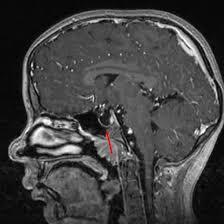
MRI Sella
Medintu has collaborated with the best pathology laboratories that are NABL and NABH certified and follow ISO safety guidelines to provide the best MRI scan for Sella at an affordable price for needy individuals. Magnetic Resonance Imaging of the sella turcica is technique used to take images of the sella turcica, which is a small bony structure within the base of the skull housing the pituitary gland. This imaging technique is essential to diagnose a variety of diseases that occur in the pituitary gland and lesion nearby structures including tumour, cysts, and inflammatory diseases. The pituitary gland, also known as the hypophysis, is the gland that directs virtually all other glands as well as miscellaneous hormones that control growth, metabolism and reproduction. Consequently, correct definition of this area is critical when diagnosing disorders that can cause hormonal disorders and other diseases. MRI is preferred in clinics due to the capability of producing the best quality image without using ionizing radiation which makes it safe for patients. Incited by headache, visual changes or hormonal disorders, MRI of the sella helps in diagnosis and successful approach to pituitary disorders.
To schedule an appointment for MRI for Sella, simply contact Medintu or call our customer care at +919100907036 or +919100907622 for more details and queries.
Overview
Specification
FAQ
Overview
What is MRI?
MRI stands for Magnetic Resonance Imaging which is a non-invasive imaging technique used to study detailed structures inside the body. This involves the use of magnetic fields and radio waves to produce pictures of body organs and tissues. MRI machines have a powerful magnetic field that attracts the hydrogen atoms in the human body, and focuses them. Radiofrequency pulses are then used that bring these atoms to signal out. These signals are then ever so slightly reformed by a computer to provide detailed cross-sectional pictures. MRI as we know is complementary to CT and X rays in imaging but with an advantage of not using radiation hence can be used often. MRI gives a great mortality in distinguishing between various types of tissues and is most beneficial for the imaging of the brain and spinal column as well as for analyzing joint conditions. MRI is used to diagnose a number of diseases and ailments, where the primary areas include Brain & spinal cord disorders, Musculoskeletal system – including tears and arthritis, Abdomen & pelvic organ disorders, Tumours and cysts. Generally safe, however, some precautions are needed for people with metallic implants or with pacemakers and other electrical devices.
Symptoms for MRI of the Sella
MRI of the sella is usually done for several purposes relating to sella turcica and pituitary gland pathology. Here are some common indications:
- Pituitary Tumors
Adenomas: Du incarceration The two most frequent benign neoplasms of the pituitary gland.
Carcinomas: Benign and malignant neoplasms that are associated with pituitary gland origin or which may involve this gland as a metastatic site.
- Cysts
Rathke’s Cleft Cysts: Benign cysts in which the tumor may have a capacity to manifest symptoms due to direct pressure.
Other Cysts: For instance, tumours such as pituitary cysts or arachnoid cysts, intracranial tumours simply mean tumours in or around the brain.
- Inflammatory Diseases
Sarcoidosis or hypophysitis the inflammation of the pituitary gland.
- Vascular Abnormalities
For example the patient had other comorbidity like pituitary apoplexy, or any other form of any vascular malformations.
- Structural Anomalies
Pathological changes in the sella or its vicinity.
- Endocrine Disorders
Assessment of conditions leading to hormonal dysfunction for example, Cushing’s disease or acromegaly.
- Neurological Symptoms
Other head pains that cannot be put down to any other cause, blurring of vision, or neurological changes that may be due to pituitary tumour.
- Follow-Up Imaging
Follow-up care of known diseases or evaluating treatment outcomes, for example, after operation, radiation therapy.
What is the Procedure for MRI Procedure?
The general technique used when carrying out an MRI of the sella is very simple in order to avoid distressing the patients while getting the best scans. Here’s an overview of what to expect:
- Preparation
Pre-Appointment Instructions: There may be some dietary restrictions as well as some specifics of the case, for example, the patient should not take some drugs in the period prior to the examination.
- Arrival and Check-In
Patients come to the imaging facility, register and some of them complete questionnaires about their condition.
- Changing into a Gown
Patients may be requested to undress partially, or in some cases wear a hospital gown because of the effects that some clothes with metallic buttons have on the machinery.
- Positioning
Patients can either sit or lie on a cradle table which is movable and for this case the patient lays flat on their back.
Sometimes the cushions or straps and other things are needed to fix the patient during the scan.
- Entering the MRI Machine
The table rolls into the MRI machine, which is a big, round pipe.
In sella imaging, the head is approached near the center of the machine, which isn’t true for other imaging techniques.
- During the Scan
Here the MRI technologist will walk out of the MRI compartment but will be able to talk to the patient over an intercom.
Patients may have some noise disturbances in the form of tapping or thumping noise produced by the machine; the patient may be provided with ear plugs or headphones to reduce noise.
- Scan Duration
The whole MRI scan can vary in time from 20 to 60 minutes depending on the protocols used and the number of sequences.
- Results
The images are read by a radiologist, the radiologist either reads the findings or dictates a report to the referring practitioner. The physician will then explain the results to the patient.
Interpretation of MRI Results
1.Normal Findings
Normal Sella Turcica Size and Shape: The sella should have an oval configuration at the bottom side and should not show any feature indicating increase in size or disintegration.
Normal Pituitary Gland: The pituitary gland should be of near normal size and location sharply demarcated from surrounding structures.
- Abnormal Findings
Pituitary Adenomas:
Description: As a rule, discrete lesions which can potentially lead to the pituitary enlargement.
Implications: They can be functioning which secrete hormones and non-functioning ones. Additional examination may be required concerning hormone balance and possible therapy.
Pituitary Carcinomas:
Description: Occasionally; may manifest as infiltrating tumours.
Implications: It may demand a more assertive approach; the patient may need operation or chemotherapy.
Cysts:
Rathke’s Cleft Cyst: Usually sits in sella, may compress the pituitary gland.
Implications: Otherwise generally innocuous, though may cause symptoms if lesions are large or if they cause symptoms themselves.
Pituitary Apoplexy:
Description: Bleeding within the pituitary gland at presentation; can have bright signals on T1 sequences.
Implications: It very often demands an emergency medical treatment.
Empty Sella Syndrome:
Description: Due to the same reason of CSF filling the sella it has the enlarged shells along with flattening of the pituitary gland.
Implications: May be without symptoms or may be accompanied by hormonal deficiencies.
Infiltrative Diseases:
Description: Sarcoidosis is another disorder which may implicate the pituitary gland, manifesting as thickening of the pituitary stalk, or as infiltrative forms.
Implications: ROC-TIME retinal oximetry has shown differences pointing at the underlying condition that needs further assessment and treatment.
- Additional Considerations
Vascular Abnormalities: Like cerebral aneurysms or other vascular abnormalities which may demand emergency treatment.
Anatomical Variations: It is also important to distinguish normal variants from pathologic findings.
Implications of Findings
MRI findings concerning the sella turcica and the pituitary gland can have major implications for the clinical course and treatment regimen of the patient. Here’s a breakdown of various findings and their potential implications:
- Pituitary Adenomas
Implications:
Functioning Adenomas: These can result in endocrine disorders such as Cushing’s disease, acromegaly, PMS and menstrual irregularities^ Hormonal disorders: These can manifest from disorders such as Cushing’s disease, acromegaly, PMS and menstrual irregularities. Treatment might be medical therapy, surgical therapy or radiation therapy.
Non-Functioning Adenomas: If asymptomatic, then is usually followed but may need surgery, where they are symptomatic for example, when they present with headaches or changes in vision.
- Pituitary Carcinomas
Implications:
These are essentially rare and aggressive malignancies that are typically managed by surgery, radiation therapy and possibly chemotherapy. The follow-up imaging should be done regularly.
- Growths
Implications:
Mainly asymptomatic, and although not usually treated, may end up being treated when they become symptomatic. Patients with large cysts which cause pressure symptoms may warrant surgical management.
- Pituitary Apoplexy
Implications:
This medical emergency cries for intervention especially by administering corticosteroids and surgical decomposition in case of neurological complications or severe manifestations of the condition.
- Empty Sella Syndrome
Implications:
May be without symptoms though can be linked to endocrine dysfunctions. For example, the endocrine assessment might be required to ensure that the patient’s pituitary gland is functioning properly and whether the patient needs to take hormonal medications.
- Granulomatous Diseases
Implications:
He or she needs to be treated for the cause, which might entail corticosteroids or other immunosuppressive drugs.
- Vascular Abnormalities
Implications:
Diseases of this type may call for surgical or even endovascular repair to prevent formation of an aneurysm which might lead to hemorrhage for instance.
- Neurological Symptoms
Implications:
Headache or change in vision might require follow-up, treatment of the conditions that cause these expressions, and referral to an ophthalmologist where surgical or medical interference is needed.
Specification
- Test Type: MRI Sella
- Preparation:
- Wear a loose-fitting cloth
- Fasting not required
- Carry Your ID Proof
- Prescription is mandatory for patients with a doctor’s sign, stamp, with DMC/HMC number; as per PC-PNDT Act
- Reports Time: With in 3-4 hours
- Test Price: Rs.3500
FAQ
What is an MRI of the sella?
MRI of Sella is an imaging procedure that provides a view of sella turcica and pituitary gland. It assists in diagnosing tumours, cysts and other formations.
What is the reason to have an MRI of the sella?
These include; those presented with new onset headache, visual complaints, other signs of endocrinopathies or in any patient where pituitary pathology is suspected.
Is the MRI procedure painful?
No, MRI procedure is not invasive and patients do experience any form of pain during the procedure. Still, there is general discomfort while on the table and one may hear some loud noise from the machine.
How long does the MRI take?
The MRI taking usually ranges between 20 to 60 minutes due to the different images taken in a single MRI process.
Should I expect something before having an MRI?
In preparation for the scan one may be required to fast and not take any food or drink several hours before the scan if a contrast agent is to be used. All of this will be determined by your healthcare provider as he or she will provide you with special instructions.
What if I am claustrophobic?
If you have any condition that may cause apprehension while in the MRI machine such as claustrophobia, tell your doctor. These are the following: choosing an open MRI or taking a medication that will make you drowsy to minimize discomfort.
Will I need a contrast agent?
Contrast may be applied using a gadolinium agent or an ioprom chemical which enhances the image. Your healthcare provider will decide whether it should be given to you depending on your medical state.
Is there any risk involved in an MRI?
MRI is relatively safe for most patients, but there could be side effects involving the contrast dyes and with the metallic hardware in the body. Pregnant patient must always disclose to her provider about any implant or medical condition.
What should I expect after the exam? Is there anything I cannot do after having the MRI scan?
Yes, most often you do not have to restrict your activities after the MRI, unless of course you were given sedation or if the doctor restricted you from any activity.
How can I book an appointment for an MRI for Sella through Medintu?
To schedule an appointment for an MRI for Sella, simply contact Medintu or call our customer care at +919100907036 or +919100907622 for more details and queries.
Why Choose Medintu for MRI Sella?
Medintu is an online medical consultant that provides home-based medical services not only in your area but also in most cities in India, including Hyderabad, Chennai, Mumbai, Kolkata, and more. We have collaborated with diagnostic centers that have the best machines and equipment to ensure you get accurate results. Medintu provides 24-hour customer service for booking the appointment of the services and guides you with instructions. Medintu also provides the best diagnostic centers at low prices. Once you receive your test results, you can easily book an appointment with our network of experienced doctors for consultation. To schedule an appointment for MRI for Sella, simply contact Medintu or call our customer care at +919100907036 or +919100907622 for more details and queries.