


Fill out form to enquire now
MRI For Scrotum
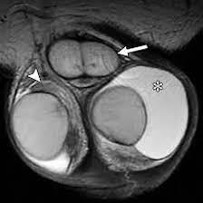
Medintu has collaborated with the best pathology laboratories that are NABL and NABH certified and follow ISO safety guidelines to provide the best MRI For Scrotum at an affordable price for needy individuals. An MRI for scrotum is a non-invasive imaging procedure that provides highly detailed, high-resolution images of the scrotal area, including the testicles, epididymis, spermatic cord, and surrounding tissues. It uses powerful magnetic fields and radio waves to create clear images of soft tissues without the need for radiation, making it a safe and effective tool for diagnosing various conditions. While other imaging techniques like ultrasound are commonly used for scrotal concerns, MRI offers superior imaging when more detailed information is needed. This can be particularly useful in diagnosing conditions such as testicular cancer, varicocele, hydrocele, epididymitis, testicular torsion, and scrotal trauma. MRI is also valuable in cases where ultrasound findings are inconclusive or when a clearer, more precise assessment is needed. MRI for scrotum is typically recommended for patients experiencing persistent pain, swelling, or discomfort in the scrotal region, or for those with suspected conditions that require further investigation. The procedure is quick, painless, and does not require the use of ionizing radiation, making it a safe and reliable option for comprehensive evaluation.
To schedule an appointment for a MRI For Scrotum, simply contact Medintu or call our customer care at +919100907036 or +919100907622 for more details and queries.
Overview
Specification
FAQ
Overview
What is MRI for Scrotum?
An MRI for scrotum is a medical imaging procedure that uses powerful magnetic fields and radio waves to create detailed images of the scrotal area, which includes the testicles, epididymis, spermatic cord, and surrounding tissues. MRI (Magnetic Resonance Imaging) provides high-resolution images that help doctors diagnose and evaluate conditions affecting the scrotum and its structures, without using ionizing radiation (like X-rays or CT scans). Unlike ultrasound, which is often the first imaging tool used for scrotal concerns, MRI offers more detailed images, especially for soft tissues, and is particularly useful in complex cases where other diagnostic methods do not provide clear information. MRI for scrotum is typically used to investigate a range of conditions, such as testicular tumors, cysts, infections, swelling, pain, or trauma in the scrotal region. It can also assist in diagnosing conditions like varicocele (enlarged veins), hydrocele (fluid-filled sac), and epididymitis (inflammation of the epididymis). The scan provides a comprehensive view of both the soft tissues and the blood vessels, helping doctors make a more accurate diagnosis and determine the best course of treatment.
Why MRI for Scrotum is Used?
An MRI for scrotum is used to evaluate and diagnose a variety of conditions affecting the scrotal area, including the testicles, epididymis, spermatic cord, and surrounding soft tissues. Here are the main reasons why MRI for scrotum is used:
- Diagnosing Testicular Tumors or Cancer
MRI is highly effective in identifying testicular tumors, helping to distinguish between benign and malignant growths. It can also determine the size and location of tumors and assess whether the cancer has spread to other areas.
- Evaluating Testicular Pain or Swelling
When a patient experiences unexplained testicular pain, swelling, or discomfort that doesn’t improve with treatment, MRI can provide more detailed information to help identify the underlying cause, such as infections, cysts, or tumors.
- Assessing Infections (Epididymitis, Orchitis)
MRI can help diagnose epididymitis (inflammation of the epididymis) or orchitis (inflammation of the testicle), which are often caused by bacterial infections. MRI can show the extent of inflammation and identify abscesses or other complications.
- Identifying Varicocele or Hydrocele
MRI is used to visualize varicocele (enlarged veins in the scrotum), which can cause pain or infertility, and hydrocele (fluid accumulation around the testicle). MRI offers a clear view of these conditions and can help determine their size and severity.
- Diagnosing Testicular Torsion
Testicular torsion, a painful and potentially life-threatening condition in which the spermatic cord twists, cutting off the blood supply to the testicle, can be difficult to diagnose. MRI is highly effective in confirming or ruling out torsion, as it provides a clear view of blood flow and tissue damage.
How MRI for Scrotum Works?
An MRI for scrotum uses magnetic fields and radio waves to create detailed images of the scrotal area, including the testicles, epididymis, spermatic cord, and surrounding tissues.
Here’s how the MRI for scrotum works:
- Magnetic Fields and Radio Waves
The MRI machine uses a powerful magnetic field to align the protons in your body, which are primarily found in water molecules.
Once the protons are aligned, radiofrequency pulses are sent into the body. These pulses temporarily disturb the alignment of the protons.
- Signal Emission and Detection
After the radiofrequency pulse is turned off, the protons return to their original alignment. As they do, they emit signals that are detected by the MRI scanner.
These signals are used to create detailed cross-sectional images (slices) of the scrotal area.
- Detailed Soft Tissue Imaging
MRI is particularly effective for imaging soft tissues, such as the testicles, epididymis, and spermatic cord, providing high-resolution images that allow doctors to assess the health of these structures in detail.
- Patient Positioning
During the MRI for scrotum, you will typically lie on your back on the MRI table. A specialized coil, which is a part of the MRI machine, may be placed near or around the scrotal area to focus the magnetic field and capture the most accurate images.
- Creating Images
The MRI machine takes multiple images (or “slices”) of the scrotum from different angles. These slices are then compiled by a computer to create a detailed, high-resolution image of the scrotal area.
- Duration of the Scan
The entire MRI scan typically lasts 30 to 45 minutes. While the procedure is taking place, you may hear loud knocking or tapping sounds as the MRI machine works. Earplugs or headphones are often provided to reduce the noise.
Benefits of MRI for Scrotum
An MRI for scrotum offers several significant advantages when diagnosing conditions affecting the scrotal area, including the testicles, epididymis, spermatic cord, and surrounding tissues. Here are the key benefits of choosing MRI for scrotal health:
- High-Resolution Imaging
MRI provides detailed, high-resolution images of soft tissues, making it an ideal tool for visualizing delicate structures in the scrotum that may not be clearly seen with other imaging techniques.
- Non-Invasive and Radiation-Free
Unlike X-rays or CT scans, MRI does not use ionizing radiation, making it a safer alternative, especially for repeated imaging or monitoring over time.
It is a non-invasive procedure that does not require surgery or needles (except for potential contrast dye administration), offering a comfortable and stress-free experience.
- Accurate Diagnosis of Complex Conditions
MRI is particularly useful for diagnosing complex conditions such as testicular cancer, testicular torsion, epididymitis, and varicocele, especially when other imaging tests (like ultrasound) have not provided clear answers.
- Enhanced Visualization of Soft Tissues
MRI provides excellent contrast between different soft tissues, allowing for detailed imaging of muscles, tendons, nerves, and blood vessels in the scrotal region. This is particularly helpful in detecting inflammatory conditions such as orchitis (inflammation of the testicle) or epididymitis (inflammation of the epididymis).
- Effective Evaluation of Testicular Torsion
Testicular torsion, a potentially serious condition where the spermatic cord twists and cuts off blood supply to the testicle, can be difficult to diagnose with ultrasound alone. MRI provides highly accurate images that can confirm or rule out torsion and help guide prompt treatment to preserve the testicle.
- Clear Detection of Scrotal Trauma or Injury
In cases of scrotal trauma or injury, MRI can help assess the extent of damage to the testicles or surrounding tissues, identifying issues such as bleeding, tearing, or swelling.
Conditions Diagnosed with MRI for Scrotum
An MRI for scrotum is a highly effective diagnostic tool for identifying a wide range of conditions affecting the scrotal area, including the testicles, epididymis, spermatic cord, and surrounding soft tissues. Here are some of the most common conditions diagnosed with an MRI for scrotum:
- Testicular Cancer
Testicular cancer is one of the most serious conditions MRI can help diagnose. MRI provides detailed imaging that helps determine the size, location, and extent of the tumor, as well as whether it has spread to other areas (metastasis).
- Varicocele
A varicocele is the enlargement of the veins within the scrotum, often causing pain and potential fertility issues. MRI can clearly visualize the size and extent of the varicocele and assess its impact on surrounding structures, including blood flow.
- Hydrocele
A hydrocele is a fluid-filled sac around the testicle that can cause swelling and discomfort. MRI can provide detailed images of the fluid collection and help differentiate hydrocele from other scrotal conditions, such as tumors or hernias.
- Epididymitis
Epididymitis is an inflammation of the epididymis, often caused by bacterial infection or trauma. MRI can accurately identify the extent of the inflammation and help determine whether an abscess or other complications are present.
It also helps in identifying chronic infections or scarring of the epididymis.
- Orchitis
Orchitis refers to inflammation of the testicle, often due to a bacterial or viral infection, such as mumps. MRI can detect the inflammation, any associated fluid collections, and other complications like abscess formation or tissue damage.
When is MRI for Scrotum Recommended?
An MRI for scrotum is recommended when a healthcare provider needs to obtain detailed, high-resolution images of the scrotal area to diagnose or evaluate various conditions. Here are the key situations where MRI for scrotum is recommended:
- Unclear Diagnosis After Ultrasound
If ultrasound results are inconclusive or if more detailed images are needed to confirm or rule out a diagnosis, an MRI may be recommended. MRI provides superior resolution and can give a clearer view of soft tissues, blood vessels, and tumors that may not be visible on ultrasound.
- Suspected Testicular Cancer
MRI is often used to evaluate testicular tumors when there is suspicion of testicular cancer. MRI provides detailed images that can help differentiate between benign and malignant masses, determine the tumor’s size, location, and extent, and check for spread to nearby lymph nodes or other tissues.
- Severe Testicular Pain or Swelling
When a patient experiences persistent testicular pain, swelling, or discomfort that doesn’t improve with treatment, MRI can help diagnose the cause, especially if conditions like epididymitis, orchitis, or hydrocele are suspected.
- Testicular Torsion
Testicular torsion is a medical emergency where the spermatic cord twists, cutting off blood supply to the testicle. If ultrasound is inconclusive, or if more detail is needed to assess the extent of the torsion and the damage to the testicle, an MRI can provide a clear view of blood flow and confirm the diagnosis.
- Scrotal Trauma or Injury
In cases of scrotal trauma, MRI is recommended to assess the extent of the injury. MRI can detect testicular rupture, hematomas (blood collections), tearing, or other internal damage caused by blunt force trauma or accidents.
- Evaluation of Varicocele
A varicocele is an enlargement of veins in the scrotum, often linked to infertility. If an ultrasound is not conclusive or if the condition is complicated, an MRI may be used to assess the size and severity of the varicocele and any impact it has on nearby structures.
Specification
- Test Type: MRI For Scrotum
- Preparation:
- Wear a loose-fitting cloth
- Fasting not required
- Carry Your ID Proof
- Prescription is mandatory for patients with a doctor’s sign, stamp, with DMC/HMC number; as per PC-PNDT Act
- Reports Time: With in 3-4 hours
- Test Price: Rs.4500
FAQ
How can I book an appointment for a MRI For Scrotum through Medintu?
To schedule an appointment for an MRI For Scrotum, simply contact Medintu or call our customer care at +919100907036 or +919100907622 for more details and queries.
What is an MRI for Scrotum?
An MRI for scrotum is a non-invasive imaging procedure used to get detailed, high-resolution images of the scrotal area, including the testicles, epididymis, spermatic cord, and surrounding tissues. MRI is often used to diagnose conditions like testicular cancer, varicocele, testicular torsion, or scrotal trauma.
Is MRI for Scrotum Safe?
Yes, MRI is generally considered safe. It does not use ionizing radiation like X-rays or CT scans, making it a safer option for imaging. However, you should inform your doctor if you have any metal implants, such as a pacemaker, as these can interfere with the MRI process. Pregnant women should also inform their doctor before scheduling an MRI.
How Should I Prepare for an MRI for Scrotum?
Wear loose, metal-free clothing and remove any jewelry, watches, or accessories before the scan. If you’re receiving contrast dye, you may need to fast for a few hours before the procedure. You should also inform your doctor of any medications or allergies you have, especially if you’re sensitive to contrast agents.
Do I Need to Fast Before the MRI?
Usually no. In most cases, you do not need to fast for a scrotal MRI unless additional tests are being performed or your doctor has specific instructions. If contrast dye is used, your doctor may recommend that you avoid eating or drinking for several hours before the scan.
What Happens During the MRI for Scrotum?
During the MRI, you will lie on your back on an MRI table. A coil may be placed around the scrotum to get detailed images. You will need to remain as still as possible for the duration of the scan, which typically takes 30-45 minutes. The MRI machine will make loud noises, but earplugs or headphones will be provided.
Does MRI for Scrotum Hurt?
No, the MRI procedure is painless. You may feel some discomfort from the position you need to maintain during the scan, but there is no pain involved. If contrast dye is used, it may be injected into a vein in your arm, which can cause a brief sensation of coolness.
How Long Does the MRI Take?
The actual MRI scan typically lasts between 30 and 45 minutes, although the entire appointment, including preparation and any paperwork, may take a bit longer.
Is Contrast Dye Used During the MRI?
Not always. Contrast dye (gadolinium) is used in some cases to enhance the images, especially when evaluating conditions like testicular cancer or vascular issues. If contrast is used, it will be injected into your arm before the scan, and you may be monitored for any allergic reactions afterward.
What Happens After the MRI for Scrotum?
After the MRI, you can usually resume normal activities immediately. If you received contrast dye, drinking plenty of water afterward can help flush it out of your system. If you were given a sedative for anxiety, you may need to arrange for someone to drive you home.
Why Choose Medintu for MRI For Scrotum?
Medintu is an online medical consultant that provides home-based medical services not only in your area but also in most cities in India, including Hyderabad, Chennai, Mumbai, Kolkata, and more. We have collaborated with diagnostic centers that have the best machines and equipment to ensure you get accurate results. Medintu provides 24-hour customer service for booking the appointment of the services and guides you with instructions. Medintu also provides the best diagnostic centers at low prices. Once you receive your test results, you can easily book an appointment with our network of experienced doctors for consultation. To schedule an appointment for an MRI For Scrotum, simply contact Medintu or call our customer care at +919100907036 or +919100907622 for more details and queries.