Fill out form to enquire now
MRI For Pelvis
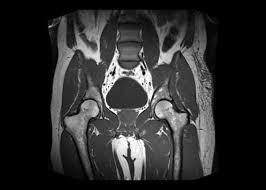
Medintu has collaborated with the best pathology laboratories that are NABL and NABH certified and follow ISO safety guidelines to provide the best MRI For Pelvis at an affordable price for needy individuals. Pelvic MRI is a powerful, non-invasive imaging technique that provides highly detailed images of the pelvic region to help doctors diagnose and monitor a wide range of conditions affecting the organs and structures in this area. Pelvic MRI uses magnetic fields and radio waves to create clear, high-resolution images of soft tissues, bones, and blood vessels without the use of radiation, making it a safe and effective tool for assessing pelvic health.
The pelvic region is the habitat of critical organs such as the bladder, reproductive organs like the uterus, ovaries, prostate, and testes, and parts of the gastrointestinal system. Thus, MRI for the pelvis is crucial in the diagnosis of uterine fibroids, ovarian cysts, prostate cancer, pelvic floor disorders, and more. It can also give very important information for staging cancers, planning surgeries, or even assessing the effectiveness of continuing treatment. To schedule an appointment for an MRI For Pelvis, simply contact Medintu or call our customer care at +919100907036 or +919100907622 for more details and queries.
How Does Pelvic MRI Work?
A Pelvic MRI (Magnetic Resonance Imaging) is a non-invasive diagnostic tool that uses strong magnetic fields and radio waves to create detailed images of the structures in the pelvic region, including the bladder, reproductive organs, and pelvic bones. Here’s an overview of how a pelvic MRI works and what it helps to diagnose.
- The Basics of How MRI Works:
Magnetic Field and Radio Waves:
Magnetic Field: MRI machines have a strong magnetic field that aligns the protons in the body with the magnetic field.
Radiofrequency Pulses: Once the protons are aligned, the MRI machine sends radiofrequency pulses into the body. These pulses “flip” the protons out of alignment.
Signal Transmission: Immediately after the RF pulse terminates, protons start getting realigned. In doing this, energy is released through electromagnetic waves.
Signal Receving: Electromagnetic waves are sensed by coils wrapped over the organ under study. Information is generated and processed inside the MRI and forms detailed pictures of the anatomy inside the human body
- Sequences used for Imaging MRI
Different sequences of MRI are often used in pelvic MRI to capture certain types of information, such as:
T1-Weighted Imaging: This imaging provides high-resolution images that allow better visualization of anatomical structures. It is very useful for evaluating the fat and water content of tissues.
T2-Weighted Imaging: T2 imaging produces greater contrast between different tissues, making it useful in outlining inflammation, fluid accumulation, or other masses.
DWI is used to measure the diffusion of water molecules in tissue and can help identify cancer or other pathology.
Post-contrast imaging: The MRI scan may involve administering a contrast agent, which is usually based on gadolinium, intravenous, into the bloodstream during the scan. This helps illuminate specific areas, such as tumors, inflammation, or blood vessels.
- What a Pelvic MRI Can Evaluate
Pelvic MRI diagnoses numerous organs and tissues found within the pelvis; valuable information for a diagnosis over several conditions, such as the following:
Female Pelvic MRI
Uterus and Ovaries:
Fibroids: MRI is very effective for the identification and characterization of size, location, and type of fibroids.
Endometriosis: MRI can identify endometrial tissue outside the uterus and commonly in the ovaries or pelvic walls.
Ovarian Cysts and Tumors: MRI may produce images that can be useful in defining cysts or masses in the ovaries, helping distinguish between benign and malignant growths.
Pelvic Inflammatory Disease (PID): MRI may reveal signs of infection or inflammation in the reproductive organs.
Adenomyosis: MRI can visualize the thickening of the uterine walls, which characterizes adenomyosis, which is a condition in which the tissue lining the uterus grows into the muscle wall of the uterus.
- Pelvic MRI for Inflammation and Infection:
Infections: MRI is highly sensitive for detecting infections or abscesses in the pelvic organs, which include the bladder, prostate, uterus, ovaries, and surrounding soft tissues.
Inflammatory Diseases: Conditions like Crohn’s disease and ulcerative colitis might involve the pelvic area; MRI evaluation may be used in assessing inflammation or complications.
What Conditions Can Pelvic MRI Diagnose?
A Pelvic MRI is a highly versatile imaging tool that provides detailed pictures of the organs, soft tissues, and bones in the pelvic region. Here’s an overview of the main conditions that a pelvic MRI can help diagnose:
- Female Pelvic Conditions:
Reproductive Organs:
Uterine Leiomyomas: Fibroids. MRI is the gold standard for the assessment of the size, location, and number of fibroids, which constitute benign tumors of the uterus. MRI may also help in differentiating between types of fibroids.
Endometriosis: MRI can identify endometrial tissue growing outside the uterus, often involving the ovaries, fallopian tubes, or pelvic walls. It is particularly useful in identifying deep infiltrative endometriosis, which is hard to diagnose with other imaging modalities.
Adenomyosis: MRI is very sensitive for adenomyosis, a condition in which the endometrial tissue (lining of the uterus) grows into the muscular wall of the uterus, causing an enlarged uterus and heavy menstrual bleeding.
Ovarian Cysts and Tumors: MRI can identify cysts or masses on the ovaries and help differentiate benign from malignant lesions. It provides detailed imaging of the ovaries and surrounding tissues.
Ovarian Cancer: MRI, especially with contrast, is useful in the detection of ovarian cancer, staging, and evaluation of the spread to surrounding tissues.
- Male Pelvic Conditions:
Prostate:
Prostate Cancer: Multiparametric MRI (mpMRI) is the mainstay imaging used in the detection and staging of prostate cancer. It can identify suspicious areas in the prostate that may need biopsy and determine the extent of the cancer.
BENIGN PROSTATIC HYPERPLASIA (BPH). MRI can evaluate the prostatic size and any changes resulting in obstructive symptoms including urinary retention or urgency.
PROSTATITIS (PROSTATE INFLAMMATION OR INFECTIOUS PROCESS). MR imaging is helpful for noting inflammation or infection of the gland, including abscess development.
TESTICULAR AND SCROTAL DISEASES
TESTICULAR CANCER. M R imaging is employed when staging testicular masses and ruling out involvement of adjacent anatomical structures.
- Conditions Involving the Pelvic Bones and Soft Tissues:
Pelvic Fractures: MRI is very good at identifying pelvic bone fractures that may not be seen on X-ray or any related soft tissue injury.
Soft Tissue Tumors: MRI is very sensitive in the identification of soft tissue tumors within the pelvis, which can involve muscles, fat, and other connective tissues, like sarcomas.
- Gastrointestinal and Other Abdominal Conditions:
Crohn’s Disease: MRI can be applied to outline the extent of Crohn’s disease in the gastrointestinal tract with regard to pelvic and rectal involvement. It is quite useful in identifying fistulas, strictures, and inflammation.
Ulcerative Colitis: MRI sometimes may be used to ascertain the degree of inflammation that might be present in the colon, especially when complications, such as perforations and abscesses, may be suspected.
Rectal Cancer: MRI is employed for the assessment of rectal tumors, staging, and the spread of the disease to adjacent structures.
- Pelvic Vascular Conditions:
Aneurysms: MRI can detect aneurysms of the pelvic arteries, including the aortic aneurysm that extends into the pelvic region, or aneurysms of the iliac arteries.
Pelvic Venous Disorders: MRI can aid in assessment of pelvic congestion syndrome, with varicose veins involving the pelvic region, particularly in females, thereby causing pelvic pain and discomfort.
Benefits of Pelvic MRI
A Pelvic MRI (Magnetic Resonance Imaging) provides numerous advantages in diagnosing and assessing a variety of conditions within the pelvic region. Here’s an overview of the key advantages of a pelvic MRI:
- High-Resolution Imaging
Clear Visualization of Soft Tissues: Pelvic MRI is especially useful in visualizing soft tissues, such as the uterus, ovaries, prostate, bladder, rectum, and muscles in the pelvis.
Superior to Other Imaging Modalities: Compared to ultrasound and CT scans, MRI provides superior soft tissue resolution, making it ideal for detecting tumors, cysts, and inflammation in the pelvic organs.
- Non-Invasive and No Radiation Exposure
No Ionizing Radiation: Unlike CT scans and X-rays, MRI does not involve exposure to ionizing radiation, which can be a concern, especially for repeated imaging or for younger patients.
Non-Invasive: MRI does not require surgery or invasive procedures to obtain detailed images of the pelvic region, reducing patient discomfort and risk of complications associated with invasive diagnostic techniques.
- Comprehensive Evaluation of Pelvic Structures
Evaluation of Multiple Organs: MRI can simultaneously assess a variety of pelvic structures, including the bladder, reproductive organs (uterus, ovaries, prostate), pelvic bones, and soft tissues.
Multiplanar Imaging: MRI provides images in multiple planes, namely axial, sagittal, and coronal, and thereby gives a 3D view of the area so that physicians can assess the location, size, and relationship between different anatomical structures better.
- Accurate Diagnosis and Disease Staging
Detection of Tumors and Cancers: MRI is very sensitive in detecting pelvic cancers, including ovarian, uterine, cervical, prostate, and rectal cancer. It is also useful in the staging of cancer, which describes the extent of the tumor and its spread to surrounding tissues.
Soft Tissue Tumors and Masses: MRI best detects benign and malignant soft tissues in the pelvis region. Whether the pathology could be a uterine fibroid, ovarian cyst, or any form of pelvic sarcoma.
Finding Metastasis: Through MRI, pelvic cancerous diseases can extend their tumors to lymph nodes, bone, or to any part of the structure adjacent to it.
- Assessment of Inflammatory and Infectious Conditions
Pelvic Inflammatory Disease (PID): MRI is quite sensitive to PID, an infection with inflammation of the female reproductive organs.
Endometriosis: MRI is regarded as one of the best imaging techniques to diagnose endometriosis, particularly deep infiltrative lesions that might involve the ovaries, pelvic peritoneum, or bowel.
Adenomyosis: MRI is very sensitive to the diagnosis of adenomyosis, which is the infiltration of the uterine lining into the muscular wall of the uterus.
Preparing for Pelvic MRI
Preparing for a pelvic MRI requires just a few important steps that ensure it goes smoothly and allows for getting the best possible images. This is what you need to know to prepare for your pelvic MRI:
- General Preparation Instructions
Clothing:
Wear comfortable clothing free of any metal parts. Examples include zippers, buttons, buckles, or snaps. In case of clothing with metal components, or if you cannot take off these clothes, you are likely asked to change into a hospital gown.
Avoid wearing jewelry, watches, hairpins, piercings, or any other metal accessories, as metal objects can interfere with the MRI’s magnetic field and distort the images.
Remove Metal Objects:
Metal can interfere with the MRI scan. Be sure to remove any items such as:
Jewelry, Watches, Hairpins, bobby pins, or other metallic hair accessories.
- Eating and Drinking
Contrast MRI: If your MRI scan requires a contrast agent, commonly gadolinium-based, you may be advised to fast for 4-6 hours before the procedure so that the risk of experiencing nausea or other side effects from the contrast agent is avoided. However, this may be different based on the instructions issued by your healthcare provider or the MRI center.
- Full Bladder Requirement
For some pelvic MRIs, for example, those assessing the bladder, uterus, or prostate, you may be asked to arrive at the exam with a full bladder to better visualize the pelvic organs.
How to Prepare: You will be asked to drink a specified amount of water (usually around 24-32 ounces, or about 700-950 mL) about 1 hour before the exam and avoid urination until after the scan.
- Medication Use
Inform the MRI staff about your medications: Make sure you tell your doctor or MRI technologist about any medications you are on, including over-the-counter medications, herbal supplements, and any prescription medications.
Inform them of any allergies to medications or contrast agents like gadolinium.
- Come Early
Arrive 15-30 minutes before your scheduled time for your MRI. This will enable you to fill out any required paperwork, review your medical history, and prepare for the actual scan. It will also give you a few minutes to ask last-minute questions or voice your concerns.
- Expectations of Pelvic MRI
The Procedure: For the MRI scan, you will be asked to lie down on the MRI table. This table will move into the narrow MRI machine (the “tube”). During the scan, the machine creates loud, rhythmic noises. You may be given earplugs or headphones to reduce the noise.
Scan Time: A pelvic MRI usually takes about 30-60 minutes, depending on the complexity of the imaging.
- Test Type: MRI For Pelvis
- Preparation:
- Wear a loose-fitting cloth
- You will be asked to fast for 4 hours prior to your exam
- Carry Your ID Proof
- Prescription is mandatory for patients with a doctor’s sign, stamp, with DMC/HMC number; as per PC-PNDT Act
- Reports Time: With in 4-6hours
- Test Price: Rs.4500
How can I book an appointment for an MRI For Pelvis through Medintu?
To schedule an appointment for an MRI For Pelvis, simply contact Medintu or call our customer care at +919100907036 or +919100907622 for more details and queries.
Is Pelvic MRI Safe?
Yes, Pelvic MRI is generally very safe. Since it uses magnetic fields and radio waves to generate images, there is no radiation involved, so it is safer than CT scans and X-rays. However, the patients with some implants, including pacemakers, metal prosthetics, or certain types of clips, have to avoid MRI or consult their healthcare provider about possible alternatives. You must inform your physician about any implants or devices within your body before getting an MRI.
Is Pelvic MRI Painful?
No, a Pelvic MRI is not painful. You’ll be asked to lie flat inside the MRI machine while the images are being taken. The machine will make noises, but you will be given ear protection to block out much of that noise. Some patients find lying on the MRI table uncomfortable; however, the scan itself does not cause pain.
How Long Does a Pelvic MRI Take?
This type of pelvic MRI usually lasts for 30 to 60 minutes depending on what you really want to achieve and if there will be the need for contrast agents. If the images or scan is quite detailed, the test may be longer. You will know beforehand how long it takes from your health provider.
Am I Allowed to Eat and Drink Before My Pelvic MRI?
In most cases, you will be asked to abstain from eating or drinking for a few hours before the scan. This is especially important if you are having a contrast-enhanced MRI, as it may affect the imaging quality. Your healthcare provider will give you specific instructions based on the type of MRI you are having.
What Happens During a Pelvic MRI?
Throughout the scan, you’ll be on a table which is fed inside into an MRI scanner; so, you will be advised not to move as moving around means improper images. Also, note that the machine is loud thumping, though earplugs or headphones will be provided for protection against that kind of noise. This is indicated by a possible injection of contrast dye to highlight certain regions of the pelvic area. The scanning usually takes a period of 30-60 minutes and during this process, you will be under observation at all times.
Are There Any Risks with Pelvic MRI?
For the majority of patients, it’s safe to get an MRI inside their pelvic cavities. Unless you have metallic implants like an auricle implant, a cochlear implant, aneurysm clips, or other contrabands, you’re definitely out of the eligibility circle. Some patients have usually encountered minimal reactions from the use of this dye, which causes feelings of warmth and gives metallic mouth taste. Before, however, inform your physician in case you are also with a history of kidneys.
Can I Have a Pelvic MRI if I’m Pregnant?
MRI is generally safe for pregnant women, especially after the first trimester since it does not use radiation. Unless it is medically necessary, your doctor may advise you to delay the MRI until after the pregnancy, at least in the early stage. Discuss your pregnancy before any imaging procedure with your doctor.
Why Choose Medintu for MRI For Pelvis?
Medintu is an online medical consultant that provides home-based medical services not only in your area but also in most cities in India, including Hyderabad, Chennai, Mumbai, Kolkata, and more. We have collaborated with diagnostic centers that have the best machines and equipment to ensure you get accurate results. Medintu provides 24-hour customer service for booking the appointment of the services and guides you with instructions. Medintu also provides the best diagnostic centers at low prices. Once you receive your test results, you can easily book an appointment with our network of experienced doctors for consultation. To schedule an appointment for an MRI For Pelvis, simply contact Medintu or call our customer care at +919100907036 or +919100907622 for more details and queries.


