


Fill out form to enquire now
MRI For Cranium
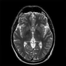
Medintu has collaborated with the best pathology laboratories that are NABL and NABH certified and follow ISO safety guidelines to provide the best MRI For Cranium at an affordable price for needy individuals. MRI of the cranium is an innovative non-invasive method of revealing brain, skull and neighbouring tissues pathology. MRI does not use radiation to take images of the brain; it employs an immensely strong magnetic field compounded by radio waves to provide multiple cross-sectional images of the area of the brain required. This makes it one of the safest and most effective of the imaging techniques that are currently in use in various departments. MRI or magnetic resonance imaging of the cranium is very important in the diagnosis of various neurological and cranial ailments such as tumours, stroke, head injuries, infections, and degenerative diseases of the brain.
It is incredibly useful for not only the initial identification of behaviours as pathological but also for the identification of the effectiveness of treatments through time. Due to the ability to get high-quality pictures of the soft tissues, MRI helps to determine the condition of the brain tissues and detect the problems in their structure that cannot be identified using another imaging method. For assessment, diagnosis, planning treatment and for follow up of patients’ conditions, MRI gives clinicians a detailed and accurate view of the state of the brain, which is crucial for decision making. Because it is not invasive and can provide rich volumetric data about the state of a patient’s head, it is an indispensable tool in contemporary medicine. To schedule an appointment for a MRI For Cranium, simply contact Medintu or call our customer care at +919100907036 or +919100907622 for more details and queries.
Overview
Specification
FAQ
Overview
Why MRI for Cranium?
Magnetic Resonance Imaging is a modern helpful method of diagnostic imaging in healthcare, especially for imaging the cranium and tissues around it, including the brain and the skull. Here are some key reasons why MRI is important for the cranium:
- Non-Invasive and Safe Imaging
As for MRI it is a non-surgical procedure so there are no cuts or injections to be made. This makes it a safer solution to some of the more invasive techniques used in diagnosing ailments such as surgery or biopsy. It is also non-ionization which makes it favorable for use compared to other imaging tests like the CT scans or X rays.
- Clear-Tissue Images
MRI offers outstanding portrayal of gentle tissues such as the brain and the nerves and blood vessels, and these are all least clear in x-ray or CT scans. This in turn helps doctors diagnose conditions of the head including the brain and skull in much better detail.
- Identifies Several Forms of Cranial Diseases
MRI is instrumental in diagnosing various conditions affecting the cranium, such as:
Brain Tumors: MRI is useful in tumor detection and mapping of the tumor as well whether malignant or benign.
Traumatic Brain Injuries: MRI is used to diagnose brain haemorrhage, oedema or any injured tissue following head injury.
Stroke: MRI can detect parts of the brain that have been damaged by a stroke and revealed early signs of the disease.
Neurological Disorders: Some of the chronic diseases, such as multiple sclerosis, epilepsy, Parkinson diseases, can be assessed for alterations of the structures.
Brain Infections: MRI is useful in diagnosing infections like an abscess or encephalitis.
- Early Detection and Diagnosis
MRI can help diagnose structural problems in the brain and skull, and the structures that are connected to them at a very early stage. Early modification and earlier diagnosis can therefore be affected thus enhancing the treatment and minimizing harm to the patients. It is important that the earliest sign is embraced as an indication of brain tumor, degenerative disease or a brain injury so that this could be an early hint for a probable successful healing.
- Guides Treatment Plans
When there are any irregularities found through MRI, then the doctors can easily make a proper and precise diagnosis. For instance, when a patient is diagnosed with a brain tumour MRI defines the size, position of the tumour and how it will affect other structures in the skull. For decisions on surgery, radiation or other treatment methods it is of vital importance to have this information.
How Does MRI for Cranium Work?
MRI for the head is a highly developed, non-invasive imaging technique that provides pictures of the brain, skull and other structures, by using powerful magnets, radio waves, and the latest computer technology. Here’s a step-by-step explanation of how MRI works for the cranium:
- Magnetic fields and hydrogen atoms
Water makes up most of the parts of the body and hydrogen is an atom that is found in the body. Such hydrogen atoms are known to be highly sensitive to the magnetic fields.
When you are tested using an MRI, you find yourself facing a powerful magnetic field of 1.5 to 3 Tesla in the MRI machine. In practice it lines up the hydrogen atoms in your body, in the brain and the tissue nearby, with the magnetic field of the MRI scanner.
- Radio Frequency Pulses
When the hydrogen atoms are lined up, the MRI machine puts a brief burst of radiofrequency (RF) energy into your body. This pulse “interferes” with the positioning of the hydrogen atoms that absorb energy and thereby positions itself outside the established magnetic field.
- Detection of Radio Waves
It works by guiding a stream of radio waves towards the body, which the MRI machine’s sensors, known as coils, pick up as the hydrogen atoms reorganize themselves. The type, the amount, and the timing of the radio waves emitted give precise characteristics of the environment around each of the hydrogen atoms.
- Image Creation
Given this data obtained from radio waves, a computer works on the signals and produces images that are cross-sectional or sections of the brain. Obtained images can be accumulated one above the other to make the three-dimensional model of the brain and all structures about it.
MRI is particularly useful when doctors need to view small areas of the brain and is far more refined than an X-ray or even a CT scan.
- Changing MRI Sequences for Certain Information
MRI technology employs varying “sequences”, or scan programs to add contrasting characteristics in the brain and skull. These sequences are designed to emphasize different tissue characteristics, such as:
T1-weighted images: And gives a clear picture of the anatomy and is frequently used to study the structure of the brain.
T2-weighted images: These are useful in revealing irregularities like tumors, inflammation or lesions. This is because those areas that are likely to have high amounts of water content like tumors or cystic lesions.
Conditions Detected by MRI for the Cranium
MRI of the cranium is a highly effective method of diagnosing a vast number of pathologies located in the brain, skull, and adjacent tissues. Below are some of the key conditions that can be detected by MRI for the cranium:
- Brain Tumors
Primary Brain Tumors: MRI can identify both intracranial tumours that are benign and malignant ones. This technique gives doctors precise pictures to identify the nature, size, position and location of the tumor, which is essential for mapping out the strategies of, for instance, surgery, radiation therapy or chemotherapy.
Metastatic Tumors: MRI is also employed to initiate the detection of metastases: tumours that originated from other body parts that have spread to the brain, determine the extent of cancer spread.
c Brain Injury (TBI)
Contusions (Bruises): Situations where MRI is useful involve confusions or hemorrhage or parts of the brain after one got involved in a head accident. MRI is especially more sensitive than CT scans because it can reveal several small areas of injury and specialize in diagnosing damages on the soft tissues.
Diffuse Axonal Injury: MRI is highly sensitive in diagnosing DAI which is a brain injury that occurs through tearing of nerve fibers usually associated with serious head trauma or an auto accident.
- Stroke
Ischemic Stroke: MRI can point to structures of the brain which do not receive blood supply because a vessel supplying the area is blocked. It serves in determination of ischemic strokes and the evaluation of the degree of tissue injury.
Hemorrhagic Stroke: MRI also works well in diagnosing hemorrhagic stroke, this is when a blood vessel breaks or bursts. This issue is essential to avoid choosing an improper emergency management mode.
- Multiple Sclerosis (MS)
MRI is the best imaging modality for diagnosing multiple sclerosis, an autoimmune condition in which the body’s defense system targets and destroys the protective sheath of nerves in the brain and spinal cord. MRI can reveal the feature of multis : periventricular regions of altered signal intensity in the white matter of the brain which is typical of multiple sclerosis.
It is also applied to evaluate the progress of the disease and to consider the effectiveness of the treatment.
- Hydrocephalus
In hydrocephalus, the normal clear fluid called the cerebrospinal fluid- CSF has collected in the ventricles of the brain in excess of the normal limits and if left untreated can cause raised pressure in the brain leading to damage of the tissues. The size of the ventricles increases, which is important for diagnosis and treatment, usually surgical, to redirect the fluid.
Benefits of MRI for Cranium
MRI for the cranium offers several significant advantages, making it a preferred choice for diagnosing brain and skull conditions:
- No Radiation Exposure: As for the MRI the conscience is less dangerous than the X-rays and computer tomography because it uses the magnet and radio waves for the repetitions.
- High-Resolution Images: MRI produces excellent images of soft tissues like the brain, nerves, blood vessels and it is useful in differential diagnosis of conditions like tumors, stroke and neurological disorders.
- Non-Invasive: MRI as a technique does not involve the use of scalpel or needles (the agents for contrast may be used sometimes though sparingly).
- Early Detection of Abnormalities: MRI enables early diagnosis of illnesses including brain tumours, strokes, and diseases affecting the nervous systems.
- Detailed Soft Tissue Visualization: In particular MRI is very useful to show kinds of soft tissues in the body including the brain’s gray and white matter, cerebrospinal fluids, and blood vessels which cannot be seen clearly through conventional x-rays or CT scans.
- Versatility: MRI can be made specific to certain sequences that allow the visualization of different tissues or pathologies and gives complete evaluation of any cranial involvement.
- Guides Treatment Plans: These pictures provide doctors with the shape, position, and specificity of the problems in a brain and assist in surgical or other therapies preparation.
- Monitoring Disease Progression: MRI can be used when diagnosing diseases such as multiple sclerosis and as a tool for observing the improvement or worsening of a disease, especially post-brain surgery or in cases of injury.
Specification
- Test Type: MRI For Cranium
- Preparation:
- Wear a loose-fitting cloth
- Fasting not required
- Carry Your ID Proof
- Prescription is mandatory for patients with a doctor’s sign, stamp, with DMC/HMC number; as per PC-PNDT Act
- Reports Time: With in 4-6 hours
- Test Price: Rs.4000
FAQ
How can I book an appointment for a MRI For Cranium through Medintu?
To schedule an appointment for a MRI For Cranium, simply contact Medintu or call our customer care at +919100907036 or +919100907622 for more details and queries.
What is an MRI of the head?
Craniotomy is a brain surgery in which a bone flap of the skull is temporarily removed to access and treat a specific area of the brain and can be paired with a procedure called an MRI for the cranium, an imaging scan for the brain and skull that provides detailed tissue data. It produces these images with the help of strong magnetic fields and radio waves which makes it possible for doctors to diagnose and observe certain neurological disorders without having to undertake radiation.
When would I need an MRI of my brain and skull?
MRI of the cranium is a diagnostic measure for various diseases including brain tumor, stroke, head injury, multiple sclerosis, infection, epilepsy, and congenital brain diseases. This is also employed in condition follow and to thereby make treatment-related decisions.
Is an MRI of the brain safe?
Yes, MRI is very safe for the most part of its usage. This one is not invasive and does not utilize radiation as the x-rays and CT scans would use. Nevertheless, some metallic implants in a patient’s body like pacemakers, metal clips or cochlear implants can make MRI difficult therefore it is advisable to inform your healthcare provider about such implants.
Does an MRI hurt?
No, an MRI is not painful. Though you will be subjected to stillness in most cases for about 20 to 60 minutes, you may experience some arils from the whole process. The machine can also make loud noises when working through the scan so you might be provided with ear plugs or headphones.
What should I do before having an MRI of the cranium?
You will probably be told to take off any items such as rings, necklaces, watches, hair clips or clothing with zippers, buttons, belt buckles etc. Before going in for the session, let your doctor know if you have any implants/ medical devices i.e. pacemaker, stent, cochlear implant, etc. If you plan on undergoing a contrast-enhanced MRI, then you could be told not to take any fluids or meals several hours prior the scan.
How long does an MRI of the brain take?
This particular scan generally lasts between 30 to 60 minutes though time varies with the kind of MRI scan that is being conducted. This will take slightly longer if contrast agents have been used.
What should I expect during the MRI procedure?
In the procedure, you are placed on a couch that is moved into a huge circular like structure. Several precautions remain necessary to guarantee good image quality: they must remain as immobile as possible. It will produce noises which are repeated throughout the scanning process and that is perfectly natural for an MRI machine. You may also be provided with ear plugs or headphones to minimize the sounds heard.
Why Choose Medintu for MRI For Cranium?
Medintu is an online medical consultant that provides home-based medical services not only in your area but also in most cities in India, including Hyderabad, Chennai, Mumbai, Kolkata, and more. We have collaborated with diagnostic centers that have the best machines and equipment to ensure you get accurate results. Medintu provides 24-hour customer service for booking the appointment of the services and guides you with instructions. Medintu also provides the best diagnostic centers at low prices. Once you receive your test results, you can easily book an appointment with our network of experienced doctors for consultation. To schedule an appointment for a MRI For Cranium, simply contact Medintu or call our customer care at +919100907036 or +919100907622 for more details and queries.