


Fill out form to enquire now
MRI For Brain Perfusion
Medintu has collaborated with the best pathology laboratories that are NABL and NABH certified and follow ISO safety guidelines to provide the best MRI For Brain Perfusion at an affordable price for needy individuals. MRI for brain perfusion is an advanced imaging technique used to assess the blood flow to different areas of the brain. This specialized MRI provides detailed, real-time information about how well blood is circulating in the brain’s tissues, which is essential for diagnosing and managing various neurological conditions. Unlike standard MRI, which focuses on the brain’s structure, brain perfusion MRI offers a unique look at the brain’s vascular system and helps identify areas where blood flow may be compromised. The procedure is primarily used to detect conditions like stroke, brain tumors, vascular malformations, and chronic cerebrovascular diseases. It can also be crucial for treatment planning, monitoring post-surgery recovery, and assessing the brain’s response to therapies such as chemotherapy or radiation. As a non-invasive, radiation-free procedure, MRI for brain perfusion is a safe and effective diagnostic tool. It plays a vital role in early detection, helping doctors make more informed decisions about treatment and care. Whether you’re undergoing the procedure to assess symptoms like dizziness or cognitive decline, or as part of a stroke evaluation, understanding what MRI for brain perfusion is and how it works can help you feel more comfortable with the process.
To schedule an appointment for a MRI For Brain Perfusion, simply contact Medintu or call our customer care at +919100907036 or +919100907622 for more details and queries.
Overview
Specification
FAQ
Overview
How MRI for Brain Perfusion Works?
MRI for brain perfusion is a specialized imaging technique that assesses how well blood is flowing through the brain’s blood vessels and tissues. Here’s how it works, step by step:
- Magnetic Resonance Imaging (MRI) Technology
Like a regular MRI, MRI for brain perfusion uses powerful magnetic fields and radio waves to generate images of the brain. The magnetic field aligns the protons in the body’s tissues, and radio waves are used to produce detailed images of the structures within the brain.
- Contrast Agent (Gadolinium) Injection
To enhance the visibility of blood flow, a contrast agent is injected into the bloodstream via a vein in the arm. This contrast dye helps highlight the movement of blood through the brain by affecting the magnetic properties of blood vessels.
- Imaging Blood Flow
After the contrast is injected, the MRI machine quickly takes a series of images of the brain over a short period. These images capture how the contrast travels through the brain’s blood vessels and into the brain tissues.
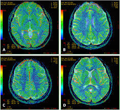
- Perfusion Mapping
The MRI machine uses advanced imaging techniques to map the perfusion—the flow of blood—across different regions of the brain. This information is processed to create perfusion maps, which show areas with normal, reduced, or absent blood flow.
- Analysis of Brain Function and Abnormalities
The resulting images and maps help doctors assess how well blood is reaching various areas of the brain. This is important for identifying brain tissue at risk or assessing brain tumor blood supply.
MRI perfusion can identify ischemic regions, and areas where brain tissue may be at risk due to vascular issues.
When is MRI for Brain Perfusion Recommended?
MRI for brain perfusion is typically recommended when there is a need to assess blood flow in the brain to diagnose or monitor certain neurological conditions. Here are some of the common situations where an MRI for brain perfusion might be recommended:
- Stroke (Ischemic or Hemorrhagic)
Ischemic Stroke: MRI for brain perfusion is commonly used in the acute phase of ischemic stroke to determine the extent of tissue damage due to lack of blood flow. It helps identify the penumbra (at-risk brain tissue) that may still be salvageable if treated promptly.
- Brain Tumors
Pre-surgical Planning: MRI for brain perfusion can help assess the blood supply to a brain tumor, which can guide surgical planning or determine the tumor’s aggressiveness.
Treatment Monitoring: It’s used to monitor the effects of chemotherapy or radiation on a brain tumor, checking for changes in blood flow to the tumor or surrounding tissue.
- Traumatic Brain Injury (TBI)
For patients with a history of head injury, MRI for brain perfusion can be used to evaluate the effects of trauma on brain tissue. It can reveal areas with decreased blood flow, helping doctors assess the severity of the injury and identify areas at risk of further damage.
- Vascular Malformations and Abnormalities
Arteriovenous Malformations (AVMs): Perfusion MRI helps assess abnormal blood flow in the brain, such as in AVMs, which are tangled masses of blood vessels where blood flow may be impaired or abnormally redirected.
- Chronic Cerebrovascular Conditions
Chronic Ischemic Disease: For individuals with long-standing cerebrovascular disease or risk factors for stroke, such as hypertension, diabetes, or high cholesterol, MRI for brain perfusion can help assess areas of chronic ischemia (lack of blood flow) and monitor the health of brain tissue over time.
Benefits of MRI for Brain Perfusion
MRI for brain perfusion offers several significant advantages in diagnosing and managing a variety of neurological conditions. Below are the key benefits of MRI for brain perfusion:
- Non-invasive and Safe
No Radiation: Unlike CT scans, MRI for brain perfusion uses magnetic fields and radio waves instead of ionizing radiation, making it a safer option for patients.
Non-invasive: The procedure does not require any surgical intervention, biopsies, or incisions, which reduces the risks typically associated with more invasive diagnostic techniques.
- Detailed Visualization of Blood Flow
Precise Mapping of Perfusion: MRI for brain perfusion creates high-resolution, detailed maps of blood flow throughout the brain, which helps identify areas with normal, reduced, or impaired circulation.
- Early Detection of Stroke and Ischemia
Ischemic Stroke: Perfusion MRI is particularly valuable in detecting ischemic strokes (where blood flow to part of the brain is blocked) by identifying areas with poor blood supply that are at risk of damage.
Acute Stroke Diagnosis: In acute stroke cases, it can determine the severity of brain damage and the potential for recovery, aiding in quicker decision-making for treatments like clot removal or thrombolysis.
- Assessment of Brain Tumors
Blood Supply to Tumors: MRI for brain perfusion helps to assess the vascularity (blood supply) of brain tumors, providing insights into whether a tumor is benign or malignant. Malignant tumors tend to have a more irregular, increased blood flow.
- Monitoring Chronic Conditions
Vascular Diseases: Perfusion MRI is valuable in monitoring chronic conditions like vascular dementia or cerebral small vessel disease, where brain blood flow is compromised over time.
Chronic Ischemia: It can track the progression of chronic ischemic disease, such as atherosclerosis or other vascular conditions, helping doctors assess the ongoing risk to brain tissue.
What to Expect During an MRI for Brain Perfusion?
Undergoing an MRI for brain perfusion is a non-invasive and generally straightforward process. However, as it involves assessing blood flow in the brain using advanced imaging techniques, there are some specific steps and preparations to be aware of. Here’s what you can expect during the procedure:
- Pre-procedure Preparation
Consultation: Before the MRI, your doctor will review your medical history, including any allergies (especially to contrast agents like gadolinium), and will ask if you have any implants, devices (e.g., pacemakers), or other conditions that might interfere with the MRI.
- Positioning on the MRI Table
Lying Down: You’ll lie on your back on the MRI table, which is padded for comfort. You will be positioned with your head in the center of the scanner. For brain perfusion scans, you may be asked to keep very still to avoid blurry images.
- Contrast Injection
Injection of Contrast: Once you are comfortably positioned, the MRI technician will inject the contrast dye (gadolinium) into your arm. The contrast helps highlight the movement of blood through the brain and allows the MRI to create detailed images of blood flow.
- The MRI Scan
The MRI Machine: The MRI machine is a large tube-shaped magnet. You will be moved into the scanner, and the procedure will typically last 30 minutes to an hour.
Sound: The MRI machine makes loud banging or thumping noises during the scan, which is completely normal. You will likely be provided with earplugs or headphones to help block the noise.
Specification
- Test Type: MRI For Brain Perfusion
- Preparation:
- Wear a loose-fitting cloth
- you may need to fast for a few hours before an MRI for brain perfusion
- Carry Your ID Proof
- Prescription is mandatory for patients with a doctor’s sign, stamp, with DMC/HMC number; as per PC-PNDT Act
- Reports Time: With in 4-6 hours
- Test Price: Rs.8000
FAQ
How can I book an appointment for a MRI For Brain Perfusion through Medintu?
To schedule an appointment for a MRI For Brain Perfusion, simply contact Medintu or call our customer care at +919100907036 or +919100907622 for more details and queries.
What is an MRI for brain perfusion?
MRI for brain perfusion is a specialized imaging technique that measures and visualizes blood flow to different regions of the brain. It helps detect abnormalities in brain circulation, which can be crucial for diagnosing conditions like stroke, brain tumors, vascular malformations, and chronic cerebrovascular diseases.
Why is an MRI for brain perfusion done?
It is done to assess blood flow in the brain, helping doctors diagnose stroke, ischemia (reduced blood flow), brain tumors, vascular malformations, and other conditions that affect brain function. It can also help monitor treatment progress or the effects of surgery, chemotherapy, or radiation.
Is MRI for brain perfusion safe?
Yes, MRI for brain perfusion is generally safe. It uses magnetic fields and radio waves, which do not involve radiation, making it safer than other imaging techniques like CT scans. However, the contrast agent used (gadolinium) may cause reactions in some people, so it’s important to inform your doctor if you have any allergies or kidney issues.
Do I need to prepare for the procedure?
Preparation may involve fasting for a few hours if contrast is used. You should avoid eating or drinking for a few hours before the MRI. Wear loose clothing without metal fasteners, as metals can interfere with the MRI. If you have metal implants, pacemakers, or other medical devices, inform your doctor before scheduling the test.
How long does an MRI for brain perfusion take?
The procedure typically takes between 30 minutes to 1 hour, depending on the complexity of the images needed and how many sequences are required.
What happens during the MRI for brain perfusion?
During the MRI, you’ll lie still on a table, and a contrast agent (gadolinium) will be injected into a vein in your arm. You will then be moved into the MRI machine, where the blood flow in your brain will be measured and visualized. The machine makes loud noises, so earplugs or headphones will be provided.
Will the contrast agent be harmful?
The contrast agent used in MRI for brain perfusion (gadolinium) is generally safe. However, some people may experience mild side effects such as a warm sensation, metallic taste, or slight nausea. In rare cases, allergic reactions may occur. If you have a history of kidney problems or contrast allergies, inform your doctor.
Does an MRI for brain perfusion hurt?
No, the MRI itself is pain-free. The only discomfort you might feel is from lying still for an extended period or the slight sting from the needle when the contrast is injected.
How should I prepare if I’m claustrophobic?
If you’re claustrophobic, let your doctor or MRI technician know ahead of time. They may offer relaxation techniques, provide medication to help you relax, or schedule the procedure in an open MRI machine (if available). You may also be able to listen to music or wear earplugs to make the experience more comfortable.
Why Choose Medintu for MRI For Brain Perfusion?
Medintu is an online medical consultant that provides home-based medical services not only in your area but also in most cities in India, including Hyderabad, Chennai, Mumbai, Kolkata, and more. We have collaborated with diagnostic centers that have the best machines and equipment to ensure you get accurate results. Medintu provides 24-hour customer service for booking the appointment of the services and guides you with instructions. Medintu also provides the best diagnostic centers at low prices. Once you receive your test results, you can easily book an appointment with our network of experienced doctors for consultation. To schedule an appointment for a MRI For Brain Perfusion, simply contact Medintu or call our customer care at +919100907036 or +919100907622 for more details and queries.