Fill out form to enquire now
CT For Sinogram
Medintu has collaborated with the best pathology laboratories that are NABL and NABH certified and follow ISO safety guidelines to provide the best CT for Sinogram at an affordable price for needy individuals. CT, a sinogram-based technique is a fundamental component of contemporary medical diagnosis and in industry, provides high-resolution, non-invasive anatomical or material analysis. This imaging technique can make high resolution cross-sectional images by converting raw X-ray projection data into sinogram for assisting doctors, researchers and engineers to take right decision. Sinogram could be defined as 2D representation of X-ray projection data, which is acquired in a number of angular positions during CT examination.
The reconstruction process enhances the sinogram from a non interpretable picture onto a recognizable image utilizing sophisticated algorithms. This process is relevant for medical diagnosis, cancer therapies, cardiovascular disease, emergency treatment and many more. Besides the healthcare domain, uses of this technology have applications in engineering, material science, archaeology, and veterinary science, where there is often a need for nondestructive testing or imaging of internal structures. To schedule an appointment for a CT for Sinogram, simply contact Medintu or call our customer care at +919100907036 or +919100907622 for more details and queries.
What is a CT Scan?
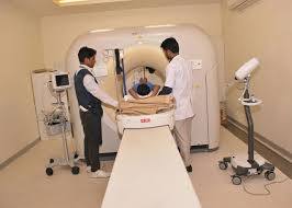
CT or computed tomography is a technique of getting images by using X-ray and computer processing to produce images in cross sectional views. While conventional examinations employ regular x-rays that deliver a two-dimensional image, CT scans offer four dimensions – a three-dimensional image. During the procedure, the patient is positioned on a table that translates into the bulk of a giant doughnut. An X-ray tube circles the body and captures pictures using different positions. A computer interprets these images and then reformats them into cross-sectional sections of the body. These slices can be superimposed on each other in order to build a 3 dimensional model. It can include: Used to Diagnose injuries for instance fracture, Detect the presence of tumors and other irregularities, Used to Evaluate diseases affecting the organs inside the body, and Help in the performance of certain treatments. They provide more detail than other imaging techniques and can identify disorders which cannot be seen through other means.
What is a Sinogram?
A sinogram is therefore a graphical presentation of the signals that are obtained through raw data from Computer Tomography Scanner scan. It has a significant function in connection with image reconstruction in CT imaging. In a CT scan, a source of X-rays revolves around the body part, and detections are taken about the projections of the X-rays. These projections indicate the density of X-rays made through the body from different orientations. The collected projection data is then reconstructed and reconstructed into a sinogram. Each point in a sinogram is the X-ray intensity sum from a given angle at a given position and captures how the body ‘casts’ a shadow of the X-ray at different angles. Columns are related to places in the X-ray trajectories.With the help of the received sinogram, it is possible to apply different complex mathematical computations and obtain cross-sectional images of the body. These reconstructions constitute the images in CT scans practiced in diagnosis. Sinograms are crucial for reconstructing the original X-ray projection data into the required 3D or 2D picture in order it could be used by doctors. That is, without sinograms ‘the data acquired by a CT scanner could not be processed as meaningful images’.
How Sinograms Work?
To better understand how sinograms work it is necessary to examine the CT imaging process and the use of sinograms in the reconstruction of images. Here’s a step-by-step breakdown of how sinograms are generated and how they work in CT imaging:
- CT Scan Data Collection
X-ray Source Rotation: In a CT scan the X-ray tube moves around the patient and takes several X-ray projections from different directions. Such projections are the actual data the scanner captures as it passes through the body with varying angles.
Detectors: Across from this is an apparatus that measures the intensity of X-rays passed through the body. These detectors determine the extent to which radiation is either absorbed or scattered as it passes through body tissues.
- Creating the Sinogram
Organizing Projections: Subsequently, the data received from the X-ray projections forms a sinogram. Projection from all angles is represented in the sinogram where each row comes from a different angle.
It has been identified that columns refer to locations along the axis on which the X-ray is taken in the body.
Each row consists of the photographs taken at a different slope of the X-ray projections in different directions.
Mathematical Transformation: The sinogram is the image of the raw data after the process has been mathematically operated on in 2D manner. This transformation tends to ‘flatten’ the analysis of the images into a grid which makes it much easier to reconstruct them back into three dimensions.
- The Sinogram’s Place in Image Reconstruction
Back-Projection: Once the sinogram is obtained, it goes through other image reconstruction techniques (FBP or iterative reconstruction etc.). These algorithms begin with sinogram data and then subserviently reconstruct the internal structure of the body in the form of cross sectional images.
Mathematics of Reconstruction: As for the sinogram data themselves this is vital as it indicates how the X-rays are affecting the structure of the body. By applying inverse operation of Radon Transform, the algorithms provide density and structural images of tissues and organs with clarity.
- Sinogram and CT Image Quality
The existence and quality of the sinogram determine the quality the reconstructed CT images. When inputting the sinogram if there is a lot of noise or mistakes in the figuring out the sinogram then there will be blurs or artifacts in the reconstructed image.
Modern generation of CT scanners and reconstruction algorithms will in one way or other try to avoid such issues by enhancing sinogram quality to produce clear images.
Significance of Sinograms in CT Imaging
In CT imaging, its function is essential for the creation of detailed cross-sectional pictures of the human body through its utilization in the construction of the high-resolution Sinograms. Here’s an explanation of its significance in CT imaging:
- Building Block for Image Construction
Raw Data Representation: It is the word used to bear the data received from the CT scanner, in a prior to processing raw format. It is a transformation of the projection data, in other words, conversion of the X-ray intensity measurements done from directions surrounding the body into another form.
Mathematical Tool: The sinogram is used in between the raw projection data and the reconstructed CT images to link the two together. The mathematical transformation such as Radon Transform performed on such raw input is enabling the generation of clear and comprehensive images of internal body structures.
- Facilitating high quality image output
Accurate Reconstructing of 3D Images: CT imaging on the other hand uses a sequence of sinograms to reconstruct the actual three dimensional images of the inside body. With the help of sinogram, by employing a high level of computer algorithms, scattered information from different projection angles can be reconstructed to create accurate two dimensional slices.
Resolution and Clarity: Bright letters were produced by high quality sinograms and this gives high resolution in images. The quality of the sinogram data is directly proportional to the resolution and contrast of the resulting CT images. For example, if the noise or some type of distortion is included in the sinogram, artifacts could appear in the reconstructed image that would locally interfere with the diagnosis.
- Improved Diagnostic Accuracy
Enhanced Visualization: Sinograms also help a clinician to view different body structures and therefore are critically important when diagnosing diseases such as tumors, bone breaks, infections or organ abnormalities. They correlate to better diaphanous images since precise sinograms indicate better pictures.
Non-invasive Imaging: To an ordinary person, it would probably be still impossible to comprehend the significance of sinograms for non-invasive diagnostics. After using mathematics to reconstruct the raw sinogram data, the doctors can see internal structures of organs, bones, and blood vessels without applying surgery and other invasive methods.
- Noise Reduction and Error Handling
Handling Artifacts: Sinograms can help identify and reduce artifacts or noise in the data, which could distort the image. For example, in the case of motion artifacts (such as patient movement during the scan), sinogram data can be adjusted to minimize these effects and improve the quality of the final CT scan.
Advanced Algorithms: Modern CT scanners use sophisticated algorithms to process sinogram data, improving the ability to handle noise and reconstruct high-quality images, even from less-than-perfect raw data.
Applications of Sinogram-based CT Imaging
- Dental Imaging
Dental CT Scans (Cone Beam CT): Sinogram-based CT imaging is used extensively in dentistry for 3D imaging of the teeth, jaw, and surrounding structures. This helps in planning dental implants, detecting cavities, or evaluating bone structure before orthodontic treatments.
Orthognathic Surgery: In cases where jaw surgery is needed, sinogram-based CT imaging provides detailed visualization of the bones and soft tissues, aiding surgeons in planning corrective surgeries.
- Musculoskeletal Imaging
Joint and Soft Tissue Evaluation: CT scans help evaluate the joints, cartilage, and soft tissues surrounding bones. Sinograms aid in producing high-quality images to detect conditions such as arthritis, ligament tears, or muscle injuries.
Spinal Imaging: For spinal injuries, herniated discs, or degenerative diseases, sinogram-based CT imaging provides detailed images of the vertebrae and surrounding structures, assisting in diagnosis and surgical planning.
- Trauma and Emergency Imaging
Trauma Imaging: In emergency medicine, sinogram-based CT imaging helps assess traumatic injuries quickly and efficiently. For example, after an accident, CT scans can be used to identify internal bleeding, fractures, or organ damage, enabling rapid decision-making in emergency situations.
Stroke Diagnosis: CT imaging is used to detect brain hemorrhages or ischemic strokes (areas of brain damage due to a lack of blood supply), helping doctors make immediate treatment decisions, such as clot removal or medication administration.
- Cardiac Imaging
Coronary Artery Imaging: CT angiography (CTA), which uses sinogram-based CT imaging, allows doctors to visualize coronary arteries in great detail. This technique helps detect blockages or other cardiovascular issues, leading to more precise treatments for heart disease, such as stent placement or bypass surgery.
Cardiac Function Evaluation: Sinogram-based CT imaging is also used in cardiac CT scans to evaluate the structure and function of the heart, allowing for better management of conditions like heart failure or valvular disease.
- Non-Destructive Testing (NDT) and Material Science
Sinogram-based CT imaging is not limited to healthcare; it has significant applications in industrial and engineering fields:
Material Inspection
Quality Control: In industries such as aerospace, automotive, and manufacturing, sinogram-based CT imaging is used to inspect materials and parts for internal defects, such as cracks, voids, or porosity, without damaging the item. This is essential for ensuring the safety and durability of products.
Additive Manufacturing (3D Printing): Sinogram-based CT scans are used to examine the internal structure of 3D-printed parts, ensuring they meet quality standards and identifying any hidden defects that might affect their functionality.
- Test Type: CT For Sinogram
- Preparation:
- Wear a loose-fitting cloth
- Fasting required
- Carry Your ID Proof
- Prescription is mandatory for patients with a doctor’s sign, stamp, with DMC/HMC number; as per PC-PNDT Act
- Reports Time: With in 4-6 hours
- Test Price: Rs.3200
How can I book an appointment for a CT for Sinogram through Medintu?
To schedule an appointment for a CT for Sinogram, simply contact Medintu or call our customer care at +919100907036 or +919100907622 for more details and queries.
What is a sinogram in CT imaging?
A sinogram is a 2D representation of the raw data collected during a CT scan. It organizes the X-ray projection data from multiple angles into a format that can be used by reconstruction algorithms to create detailed cross-sectional images (CT slices) of the body.
How does a sinogram work in CT imaging?
During a CT scan, an X-ray source rotates around the body, collecting projections (images) from different angles. These projections are then transformed into a sinogram, where each row represents one projection angle, and each column represents a position along the X-ray path. This sinogram data is then used to reconstruct 3D images of the internal structures.
Why are sinograms important for CT scans?
Sinograms are essential because they serve as the raw material for reconstructing high-quality CT images. Without sinograms, the complex data captured by a CT scanner would be unintelligible, preventing the creation of detailed diagnostic images.
How does sinogram data become a CT image?
After the sinogram is created, advanced reconstruction algorithms (such as filtered back-projection) are applied to convert the sinogram data into a detailed cross-sectional image. This process effectively “reverses” the data collection process and turns the sinogram into a clear, usable image of the body or object being scanned.
What is the role of sinograms in medical diagnostics?
Sinograms are vital in medical imaging as they allow for the precise reconstruction of CT images. These images are used to diagnose various conditions, such as tumors, bone fractures, infections, or internal bleeding, enabling accurate treatment planning and medical decision-making.
Can a sinogram be used for non-medical applications?
Yes, sinogram-based CT imaging is also used in fields such as material science, archaeology, engineering, and manufacturing. It helps with non-destructive testing (NDT) of materials, inspecting the internal structures of items like airplane parts, 3D-printed objects, and fossils, without causing damage.
How does the quality of a sinogram affect the CT image?
The quality of the sinogram directly affects the clarity and accuracy of the final CT image. If the sinogram contains noise or errors (e.g., from motion artifacts or poor data collection), the reconstructed image may have artifacts, reducing its diagnostic value. High-quality sinograms lead to high-resolution, accurate CT images.
How does sinogram-based CT imaging help in cancer treatment?
Sinogram-based CT imaging allows for detailed visualization of tumors, helping doctors assess their size, location, and spread. This is crucial for planning cancer treatments like surgery, radiation therapy, or chemotherapy, as it provides a clear map of the tumor’s characteristics.
Why Choose Medintu for CT For Sinogram?
Medintu is an online medical consultant that provides home-based medical services not only in your area but also in most cities in India, including Hyderabad, Chennai, Mumbai, Kolkata, and more. We have collaborated with diagnostic centers that have the best machines and equipment to ensure you get accurate results. Medintu provides 24-hour customer service for booking the appointment of the services and guides you with instructions. Medintu also provides the best diagnostic centers at low prices. Once you receive your test results, you can easily book an appointment with our network of experienced doctors for consultation. To schedule an appointment for a CT for Sinogram, simply contact Medintu or call our customer care at +919100907036 or +919100907622 for more details and queries.


