


Fill out form to enquire now
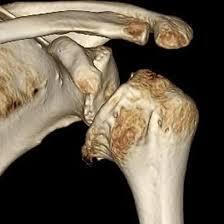
NCCT 3D for Right Shoulder
Medintu has collaborated with the best pathology laboratories that are NABL and NABH certified and follow ISO safety guidelines to provide the best NCCT 3D for Right Shoulder at an affordable price for needy individuals. Non-Contrast Computed Tomography in three dimensions (NCCT 3D) is the advanced method of shoulder imagery, which offers a precise and comprehensive evaluation of the right shoulder. NCCT 3D is a noninvasive imaging modality that employs high beam kilovoltage X-ray making it useful for diagnosing a number of shoulder pathologies based on the generation of the 3D bony images. Such a joint can be characterised as the right shoulder – in human anatomy this joint is quite cagey and requires a close attention since it may be damaged, influenced by various pathologies, and experience changes in the course of muscular dystrophy. It is also very important that imaging is achieved in the right time and most of all, accurate. Still, NCCT 3D not only helps to study the fractures, dislocations and the joint integrity, and avoid the utilisation of contrast preparations that may provoke undesirable reactions or complications. To schedule an appointment for an NCCT 3D for Right Shoulder, simply contact Medintu or call our customer care at +919100907036 or +919100907622 for more details and queries.
Overview
Specification
FAQ
Overview
What is NCCT 3D?
NCCT 3D means Non-Contrast Computed Tomography that has three dimensions. It is a technique in the diagnosis of disease through the use of X-ray technology to build up images of sections of the body without any injection of contrast medium. These scans eliminate the use of contrast materials to enhance image as widely used in the traditional computer tomography scans or CT scans instead it uses natural absorption coefficients of the X-ray through various tissues to provide 3D images. There is always an added value to enhance images of various specimens and from the NCCT, one could manipulate the images to produce end-to-end 3D images of the structures involved. With Reference to Box 3.2 this is especially useful for appraising areas that are complicated; such as joints. NCCT 3D is also applied when diagnosing bone fractures and joint dislocations as well as other orthopedic disorders. It is particularly useful when trying to obtain detailed information on areas such as the shoulder as the bony structures show up brightly. The technique shortened the time of scanning and eliminated the possibility of anaphylactic reactions or a deterioration of kidney performance caused by contrast agents for many patients.
Indications for NCCT 3D of the Right Shoulder
Here are some common indications for performing NCCT 3D imaging of the right shoulder:
- Trauma and Fractures
Evaluation of proximal humeral, scapular, and clavicular fractures.
Determining the severity of multiple traumatum which is hard to diagnose using the standard radiography.
- Dislocations
Clinical examination of any shoulder dislocation, be it anteroposterior, inferior or superior.
Fracture or ligamentous instability of the pelvic ring or involvement of the spinal column.
- Rotator Cuff Injuries
In [MeSH] Medical Subject Headings this allows much more detailed evaluation of specific bony changes associated with rotator cuff tears.
Articular-sided evaluation of matchstick-like ossifications, suggestive of impingement, or alteration in the size or shape of the greater tuberosity.
- Arthritis and joint diseases and degenerative diseases
The use of this technology for assessment of shoulder osteoarthritis, rheumatoid arthritis or other degenerative joint diseases affecting the joint.
Evaluation of joint space degeneration, periodical ossification, and other phenomena associated with arthrosis.
- Tumors and Lesions
Designation and definition of primary malignant osseous neoplasm or metastatic process at the shoulder girdle.
Diagnosis of diseases like cyst, Contusion or other lesion that do no imply bone destructions.
- Pre-Operative Planning
High resolution imaging for preoperative planning of reconstructive surgeries, for example shoulder arthroplasty or stabilisation procedures.
Evaluation of the deviations of normal anatomy that may affect the planned surgeries.
- Post-Operative Evaluation
Evaluation of the results after shoulder surgeries, fixation rigidity or union issues.
- Chronic Pain Evaluation
Evaluation of shoulder pain of uncertain nature in cases where routine diagnostic tests do not reveal the cause.
Evaluation of structural alterations secondary to chronic disorders.
The Procedure for NCCT 3D for Right Shoulder
- Preparation
Patient Assessment: A healthcare provider will need patient’s medical history and other images that the patient had before.
Clothing and Accessories: The patient might be required to undress to their surgical gown and it’s important they remove any metal on the body including jewelry, belt, etc.
- Positioning
Patient Positioning: Usually the patient will be expected to lie supine on the CT scanner table. The right shoulder will then be aligned with the right horizontal line of the scanner and the whole height focused on the shoulder.
Stabilization: A cushion or straps may be occasionally applied on the patient to avoid the patient’s movement during the scan.
- Scanning Process
Scanner Setup: Depending with the area being captured, adjustments are made on the CT machine. A positioning guide may be used by the technician to achieve proper positioning of the component.
Breath-Holding: Due to the possibility of patient movement during the scan the patient may be asked to suspend breathing momentarily.
Image Acquisition: To depict the collision zone, the scanner will swivel around the shoulder and take several axial views. On average, the process should not take more than few minutes.
- 3D Reconstruction
Image Processing: When the scan is done the images go to a computer that can further process them and make representations of the structures of the shoulder in 3 dimensions.
Analysis: Radiologists can produce different projections of the shoulder and therefore any bony structure can be well assessed for any abnormally.
- Post-Procedure Care
Recovery: There is no need for intensive care monitoring after the procedure, as NCCT does not use contrast enhancers; Most patients are then able to resume daily tasks since it does not require any intense procedure or extensive time in the operating theatre.
Results: The radiologist will interpret the plates captured and make a report to the attending doctor, who will in turn explain to the patient.
Advantages of NCCT 3D for Right Shoulder Imaging
Here are the key advantages of using NCCT 3D for imaging the right shoulder:
- High-Resolution Images
Offers pack images that make it easier to visualize small and intricate bony structures which might have been misdiagnosed.
- Visualisation in Three Dimension
Such reconstruction of images in three dimensions comes in handy when viewing the shoulder from different perspectives deepens the understanding of the image in terms of how structures are oriented.
- No Contrast Required
Compared to routine CT, NCCT 3D does not involve the use of such agents, therefore minimising the occurrence of allergic reactions as well as those pertaining the kidney function.
- Quick and Efficient
It’s common to take only some minutes for the scanning, and that is good for patients and health care workers.
- Improved Diagnostic Accuracy
Improved diagnosis of fractures dislocations and other bony lesion results in increased understanding of abnormalities and the ability to provide apt treatment for the detected conditions.
- Evaluation of Complex Cases
Especially valuable when definitive diagnosis can be made using other imaging studies such as in multifragmentary fractures or joint instability.
- Low Radiation Exposure
A new technology that is available ensures that far go odry is used that used to be used when other imaging techniques were in use hence making milder for patients.
- Chronic conditions, chronic disease assessment
Useful to assess chronic shoulder conditions like arthritis or degenerative changes in the shoulder, and gives valuable information for handling.
- Has application in medico legal cases and helps in Per and Post operative planning
Provides useful information for surgical decision making and assessment of the results, as well as improvements.
- Patient Comfort
The method is minimally invasive and therefore patients can have a good experience when receiving the procedure.
Interpreting the Results
- Initial Image Review
Radiologist Assessment: A radiologist will then evaluate the 3D reconstructed images of the shoulder to ensure that overall appearance is normal or if there is any deformity.
Comparison with Previous Studies: Depending on the Kensington current images, comparison with the previous images may enable assessment of the changes caused by the disease.
- Structural Analysis
Bony Anatomy: The radiologist will scrutinize the clavicle, scapula, humerus and all jig Glowing structures attached on the upper limb taking keen attention on establishe bony annotations.
Joint Spaces: Aspisation of joint space for any sign of joint space narrowing as evidenced by arthritic or other degenerative diseases.
- Detection of Abnormalities
Fractures: There may be difficulties in diagnosing distal radius fractures, especially fractures that might be small or complicated making them invisible on a plain X-ray.
Dislocations: Physical examination to determine if there are cases of dislocations and if there are, assessment of their damage.
Bone Lesions: Suspected tumours, cysts or any other abnormality that may need further investigation.
- Examination and interpretation of Soft Tissues (if any)
While the primary structures of interest at NCCT are bony structures, the radiologist might also look for signs of soft tissue abnormalities that might have resulted from rotator cuff pathology.
- The evaluation of related conditions
Impingement: Search for the bony spurs or changes in the acromion could be expected in the impingement syndromes.
Joint Stability: Assessment of the presence and severity of some forms or instability and other pathologies of the shoulder joint.
- Reporting Findings
Detailed Report: Besides interpretation, the radiologist will Dictate him/herself a report that will encompass the imaging study, findings, any abnormalities noted, the implications of these abnormalities, if any, management or follow-up recommendation.
Visual Aids: There should be tables when presenting the survey results, as well as graphs, annotated images or even 3D models with the description of the analyzed sections.
Specification
- Test Type: NCCT 3D for Right Shoulder
- Preparation:
- Wear a loose-fitting cloth
- Fasting not required
- Carry Your ID Proof
- Prescription is mandatory for patients with a doctor’s sign, stamp, with DMC/HMC number; as per PC-PNDT Act
- Reports Time: With in 3-4 hours
- Test Price: Rs.5000
FAQ
How can I book an appointment for a NCCT 3D for Right Shoulder through Medintu?
To schedule an appointment for a NCCT 3D for Right Shoulder, simply contact Medintu or call our customer care at +919100907036 or +919100907622 for more details and queries.
What is NCCT 3D?
NCCT 3D stands for Non-Contrast Computed Tomography in three dimensions and is a medical imaging diagnostic method where structures of the shoulder are imaged in detail, and high spatial resolution is achieved without using a contrast agent. It can provide a means of model making of bony structures for diagnostic and treatment planning purposes.
In the case of shoulder imaging why is NCCT 3D preferred?
Some benefits are the possibility to visualize complex bony structures, short scans time, non-contrast technique and still less radiation dose compared with iterative methods.
What diseases can be preliminarily identified using non-contrast computed tomography, three-dimensional?
NCCT 3D is useful in assessing fractures, dislocations, arthritis, rotator cuff injuries and tears, bone tumors and other pathology in shoulder joints.
How long does the NCCT 3D scan take?
That depends on the machine being used; it takes approximately two hours for the 3D scan to be conducted using the NCCT machine.
What special preparations are required before the scan is conducted?
In general, no preparation is required for this project. Patients should be in comfortable clothing and it is understandable that any object containing metal will have to be removed. The person should disclose any medical condition or allergy to the technician.
Is there any discomfort on the client during the procedure?
The NCCT 3D scan doesn’t cause any discomfort, and therefore is not painful at all. Sometimes patients may have to lie down for a short time, but the brief period of discomfort should not come into the picture at all.
Can there be any drawbacks linked to NCCT 3D?
The most significant risk associated with Nuclear Medicine is the exposure to radiation though through development in technology more exposure has been reduced. Concerning contrast agents there is no risk seen in NCCT because no contrast agents are used here.
What will be the steps for me to be receiving the results?
The radiologist will read the images and write a report that will be forwarded to the attending doctor. The physician will explain the results to you personally and any further action that should be taken.
Why Choose Medintu for NCCT 3D for Right Shoulder?
Medintu is an online medical consultant that provides home-based medical services not only in your area but also in most cities in India, including Hyderabad, Chennai, Mumbai, Kolkata, and more. We have collaborated with diagnostic centers that have the best machines and equipment to ensure you get accurate results. Medintu provides 24-hour customer service for booking the appointment of the services and guides you with instructions. Medintu also provides the best diagnostic centers at low prices. Once you receive your test results, you can easily book an appointment with our network of experienced doctors for consultation. To schedule an appointment for a NCCT 3D for Right Shoulder, simply contact Medintu or call our customer care at +919100907036 or +919100907622 for more details and queries.