


Fill out form to enquire now
CT scan Maxilla Axial
Are you looking for an affordable CT scan Maxilla Axial? Medintu offers competitively priced MRI and CT scans, partnering with top NABL-certified diagnostic centres and clinics. Our facilities ensure high-quality imaging and accurate results. A CT scan of the maxilla (axial view) provides detailed cross-sectional images of the upper jaw, teeth, sinuses, and surrounding structures. It is commonly used to evaluate conditions such as fractures, tumours, infections, and developmental abnormalities of the maxillary bones. The axial view captures horizontal slices of the maxilla, allowing for a clear assessment of its structure and alignment. This imaging technique is instrumental in planning dental procedures, sinus surgeries, or evaluating the extent of facial injuries. Unlike traditional X-rays, a CT scan offers more detailed, 3D images, which help doctors make more accurate diagnoses and treatment decisions. To book a Maxilla Axial CT scan appointment, visit our platform, Medintu, or contact us at +919100907036 or +919100907622 for reasonable prices.
Overview
Specification
FAQ
Overview
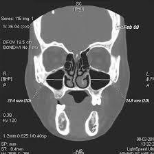
What is a CT scan of Maxilla Axial scan?
A CT scan of the maxilla (axial scan) is a specialised imaging procedure that uses computed tomography (CT) to create detailed cross-sectional (slice) images of the upper jaw (maxilla) and surrounding structures, including the sinuses, teeth, and nasal passages. The term “axial” refers to the imaging plane, which captures horizontal slices of the body from top to bottom, allowing doctors to view the area from multiple angles. This scan is handy for assessing conditions like fractures, infections, tumours, and dental abnormalities. It provides a more detailed, 3D image than traditional X-rays, helping healthcare providers make precise diagnoses and treatment plans.
Why is it Done?
A CT scan Maxilla Axial is done for various reasons:
- To evaluate fractures or trauma to the upper jaw and surrounding facial structures.
- To identify and assess the size or spread of tumours, cysts, or infections in the maxilla or sinuses.
- To check for sinusitis or other sinus-related problems affecting the maxillary sinuses.
- To assist in planning complex dental surgeries or implants by providing a detailed view of bone structure.
- To detect congenital or developmental issues with the maxillary bones, teeth, or surrounding tissues.
How to Prepare for a CT Scan Maxilla Axial.
Below are the following preparations required for a CT scan Maxilla Axial:
- Let the technician know if you have any allergies, particularly to contrast dye, or if you are pregnant or breastfeeding.
- Take off any jewellery, dentures, hearing aids, or anything else with metal, as these can interfere with the scan.
- If the scan requires a contrast dye to improve image clarity, you may be asked to refrain from eating or drinking for a few hours before the procedure.
- Opt for loose, comfortable clothing without metal parts. You may be asked to change into a gown for the scan.
- During the scan, you must lie still to ensure clear images. If you feel anxious, talk to the technician beforehand; they may offer advice to help you relax.
What is the procedure for a CT scan of the Maxilla Axial?
The procedure for a CT scan Maxilla Axial generally involves the following steps:
- You will be asked to remove any metal objects, such as jewellery, dentures, or hearing aids, and possibly change into a hospital gown to avoid interference with the scan.
- You will be on the CT table, usually lying on your back. The technician will guide you in positioning your head correctly to capture the maxilla and surrounding structures. Depending on the examined area, you may need to bite down or remain still for specific angles.
- The table will move through the CT machine, which uses X-rays to capture cross-sectional (axial) images of the maxilla and surrounding areas. The machine will rotate around your head, and you will hear a slight buzzing or clicking noise during the process.
- If contrast dye is required for better image clarity, it will be injected into a vein, usually in your arm, before or during the scan. The dye enhances the visibility of blood vessels, tissues, or abnormalities in the maxillary region.
- The scan usually takes about 10-20 minutes. Afterwards, you may be asked to wait briefly while the images are reviewed for quality. Once completed, you can return to your normal activities. The photos will be analysed by a radiologist and sent to your doctor for further evaluation.
What is the aftercare for a CT scan of Maxilla Axial?
- You can usually return to normal activities immediately after the scan unless given a sedative or contrast dye.
- If a contrast dye is injected, drink plenty of water to help flush the dye out of your system.
- While rare, some people may experience mild reactions to the contrast dye, such as a rash, itching, or a warm sensation. Contact your doctor if you experience any unusual symptoms.
- The images will be reviewed by a radiologist and sent to your doctor. You should follow up with your healthcare provider to discuss the results and any potential next steps for treatment if needed.
- There are typically no special restrictions or precautions following a CT scan unless otherwise instructed by your healthcare provider.
What is the cost of a CT scan of Maxilla Axial?
The Maxilla Axial CT scan costs Rs.2,000/-to Rs.42,000/-, and the reports are available within 24 hours, ensuring you receive your results promptly and can take the following steps in your treatment plan.
Specification
- Test Type: CT scan Maxilla Axial
- Preparation:
- Wear a loose-fitting cloth
- Fasting is required for a few hours if instructed
- Stay hydrated
- Carry Your ID Proof
- Prescription is mandatory for patients with a doctor’s sign, stamp, with DMC/HMC number; as per PC-PNDT Act
- Reports Time: With in 24 hours
- Test Price: starts from Rs.2,000/-to Rs.42,000/-
FAQ
How can I book an appointment for a low-cost CT scan of Maxilla Axial through Medintu?
Booking an appointment is simple. You can do it through our website, Medintu, by filling out the inquiry form or contacting us at +919100907036 or +919100907622. Our team will guide you through the process and ensure you get the earliest available slot that suits your schedule.
What is a CT scan Maxilla Axial scan?
A CT scan of the maxilla (axial scan) is a specialised imaging procedure that uses computed tomography (CT) to create detailed cross-sectional (slice) images of the upper jaw (maxilla) and surrounding structures, including the sinuses, teeth, and nasal passages.
What is the cost of a CT scan of Maxilla Axial?
The Maxilla Axial CT scan costs Rs.2,000/-to Rs.42,000/-, and the reports are available within 24 hours.
When will my reports be available?
Reports will be available within 24 hours after your scan.
Is a CT scan of Maxilla Axial safe?
Yes, a maxilla (axial) CT scan is generally safe, though it involves exposure to a small amount of radiation and may require a contrast dye, which can have rare side effects.
What can CT scan Maxilla Axial detect?
A maxilla (axial CT scan can detect conditions such as fractures, tumours, infections, cysts, sinus abnormalities, and other structural issues in the upper jaw and surrounding areas.
Why Choose Medintu for Your CT Scan Maxilla Axial?
Medintu is a leading digital healthcare platform that provides high-quality MRI scans for your MRI And SI Joints at affordable prices in trusted laboratories. We collaborate with top-tier labs and clinics with the latest technology to offer a wide range of ultrasound scans with accurate results. Your safety and comfort are our top priorities. We provide various ultrasounds for twins, bladder, head, knee, limbs, eyes, and more. Additionally, we offer MRI scans, such as the brain with CSF flow and lower abdomen, and CT scans like NCCT of the right foot and right tibia. Our partners are NABL and MABH certified, ensuring high-quality care. For more information, visit our page on Medintu or contact us at +919100907036 or +919100907622.