Fill out form to enquire now
CT Enterography
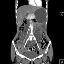
Medintu has collaborated with the best pathology laboratories that are NABL and NABH certified and follow ISO safety guidelines to provide the best CT Enterography at an affordable price for needy individuals. The following is a summary of some of the clinical indications as well as details regarding highly advanced imaging techniques including the very specialized use for enterography. CT enterography offers many benefits as this noninvasive procedure merges high resolution to CT scanning together with administration and use of specific contrast agent producing detailed images for better differentiation concerning disease processes within intestines in addition to nearby structures it presents specifically regarding diseases like IBDs/Crohn’s, small intestinal carcinomas, mechanical obstructions, and acute/ obscure melaena.
CT Enterography is not the same as other general CT scans, which are body images. It allows more vivid visualization of the small intestine than any other methods can do. It involves oral contrast, which distends the intestines, and intravenous contrast, which allows visualization of blood vessels. The technique provides a detailed view of the small bowel, so doctors can determine conditions with more accuracy, design appropriate treatments, and follow the course of the disease. CT Enterography is used in patients who require clear and precise diagnostic information but avoid more invasive procedures like endoscopy or surgery. The procedure is quick, non-invasive, and very effective in providing comprehensive insights into abdominal health. To schedule an appointment for CT Enterography, simply contact Medintu or call our customer care at +919100907036 or +919100907622 for more details and queries.
How Does CT Enterography Work?
CT Enterography works by the use of computed tomography (CT) scanning to produce high-resolution, two-dimensional images of the small intestine and the structures within it. Here’s how the test is done, step by step:
- Preparation for the Scan:
Before the CT scan, the patient must be fasting for some hours prior to the scan to empty out the small intestine of food material so that better-quality images can be taken.
Patient is told to take oral contrast around 1-2 hours before the procedure. In this process, he is required to take an oral contrast agent known as a solution. Oral contrast fills up and distends the small intestine for better visualization on a scan.
- Intravenous Contrast:
While doing the scan, an intravenous (IV) contrast agent is injected into a vein in the arm. The contrast highlights the blood vessels and tissues, so structures can be more clearly identified by taking different images that might have different appearances based on how X-rays are able to penetrate various materials.
- Procedure for CT Scan:
Patient lies down on a CT scan table. This table passes through the CT scanner, which is able to make images of the abdomen by passing X-rays through various angles.
The cross-sectional images of the small intestine and its tissues are quickly developed and subsequently transferred to the computer to reconstruct them into high resolution 2D and 3D images.
- Reconstruction
After capturing these images, a computer reconstructs them to provide sharp images of the small intestine and surrounding tissues, as well as blood vessels. All these enable doctors to visualize the presence of any abnormal changes, such as inflammation, tumors, blockage, and bleeding in the small bowel.
- Final Results:
These pictures assist doctors in diagnosing and evaluating inflammatory bowel diseases (IBD), small bowel tumors, Crohn’s disease, intestinal blockages, and other GI illnesses.
Indications for CT Enterography
CT Enterography is one of the best diagnostic techniques that have mainly been used to evaluate diseases of the small intestine and its associated abdominal structures. Some of the most common reasons for performing CT Enterography include the following:
- Inflammatory Bowel Disease (IBD):
Crohn’s Disease:
CT Enterography is very common for the diagnosis and monitoring of Crohn’s disease, one of IBD that causes inflammation in the digestive tract. It helps identify inflammatory areas, strictures, fistulas, and other complications arising from Crohn’s disease.
Ulcerative Colitis:
This includes although less commonly used to rule out ulcerative colitis is still used when Crohn’s disease and or over lay syndrome is suspected to assess inflammation grade and smaller bowel complications, where small bowel tumors are under concern.
- Small Bowel Tumors:
CT Enterography is highly useful in the diagnosis of both benign and malignant tumors of the small intestine, including adenocarcinomas, lymphomas, and neuroendocrine tumors. It has high resolution images to aid in tumor localization, size, and extent of disease.
- Small Bowel Obstruction:
CT Enterography may be useful in the diagnosis of adhesive intestinal obstructions, intestinal obstructions due to hernias, tumors, or strictures. It might help clearly outline the location and cause of obstruction and thereby guide treatment decisions.
- Gastrointestinal Bleeding:
CT Enterography is also a helpful tool when suspicion for bleeding occurs within the small intestine, especially when other methods, such as endoscopy, were unable to precisely localize the source.
- Intestinal Infections:
CT Enterography will be helpful in establishing the diagnosis of intestinal infections due to the inflammatory process caused by infections, such as bacteria and parasites. It is a platform between infectious and other gastrointestinal pathologies.
- Fistulas and Abscesses:
CT Enterography may aid in the identification of fistulas and abscesses in patients with conditions like Crohn’s disease. The identification of the collections of pus may come in handy during the planning for treatment.
Benefits of CT Enterography
CT Enterography has many advantageous uses to diagnose and manage disease in the small intestine and related structures. Here are some of the major benefits gained from using CT enterography.
- High-Resolution Images:
CT Enterography images the small intestine and surrounding tissues in high resolution. A doctor can thereby clearly visualize even the tiniest abnormality, ranging from inflammation and strictures to fistulas, tumors, and bleeding in the small intestine. Its application is necessary for all patients suspected to have symptoms of Crohn’s and other gastrointestinal illnesses.
- non-invasive.
CT Enterography is a non-invasive procedure unlike endoscopy or laparoscopy. It does not require surgical incisionation or implantation of a camera inside the body, which therefore decreases discomfort to the patient and limits the potential complications.
- Relatively Speedy and Efficient
The procedure takes 20-30 minutes to carry out, and the results are achieved within a relatively short period of time. This makes CT Enterography a time-efficient diagnostic option, especially for emergency or outpatient settings where rapid results are paramount.
- Extensive visualization of the small intestine
CT enterography is highly effective in achieving better visualization of the small intestine, which is quite hard to be assessed by other imaging techniques such as regular CT scans or X-rays. This is very useful in diagnosing diseases like IBD, small bowel tumors, and intestinal obstruction conditions.
- Assessment of surrounding structures
Besides small intestines, CT Enterography also allows an opportunity to assess the surrounding structures such as blood vessels, lymph nodes, and other adjacent organs. For this reason, doctors can obtain an overall view of the condition in the abdominal cavity and give a very detailed clinical picture.
- Better clarity with contrast enhancement
The use of both oral and intravenous contrast agents enhances visualization in the small intestine and submucosal tissues by outlining vascular structure, lesions, and inflammation, thus allowing better evaluation. The contrast helps differentiate between normal and diseased tissues.
- Test Type: CT Enterography
- Preparation:
- Wear a loose-fitting cloth
- Fasting required
- Carry Your ID Proof
- Prescription is mandatory for patients with a doctor’s sign, stamp, with DMC/HMC number; as per PC-PNDT Act
- Reports Time: With in 4-6 hours
- Test Price: Rs.4500
How to book an appointment for a CT Enterography?
To schedule an appointment for CT Enterography, simply contact Medintu or call our customer care at +919100907036 or +919100907622 for more details and queries.
What is CT Enterography?
CT Enterography is an advanced imaging that uses the CT and contrast medium to detail high-resolution images of the small intestine and the associated abdominal structure. It is performed mainly in diagnosing conditions such as Crohn’s disease, small bowel obstruction, and other intestinal tumours.
What is contrast liquid, and why am I required to consume it?
The oral contrast liquid helps to distend the small intestine, making it easier to visualize during the scan. It enhances the clarity of the images and helps differentiate the small intestine from surrounding tissues, improving diagnostic accuracy.
Is CT Enterography painful?
CT Enterography is non-invasive and relatively painless. Some patients may feel mild discomfort as a result of the contrast solution ingestion or intravenous injection of the intravenous contrast. However, this procedure is very short, and most patients tolerate it.
How long does the CT Enterography procedure take?
The whole process takes around 20-30 minutes, depending on the complexity of the scan. You have to arrive early to drink the contrast solution. Results are usually available soon after the scan is done.
Is preparation necessary for CT Enterography?
Yes, you would have to fast for some hours before the scan. Another thing is that you would have to drink an oral contrast solution before the procedure. You would be instructed to take this by your doctor; therefore, follow his instruction.
Are there any risks associated with CT Enterography?
The most important risks for these procedures are the use of the contrast agents, which occasionally result in allergic reactions that affect some patients. Again, these reactions are really infrequent and are seldom very severe. The fact that it uses radiation makes the procedures unsafe for pregnant women; any procedure involving radiation must weigh these against the benefits.
How is CT enterography different from a plain CT scan?
CT enterography allows for better definition of the images of the small intestine and their surrounding anatomical structures. It mainly incorporates oral and intravenous contrast agents for the higher resolution of images provided, which carries much more information than that done by a common CT scan.
Can the test detect the presence of Crohn’s disease?
CT Enterography is very valuable in diagnosing Crohn’s disease. It depicts inflammation, strictures, fistulas, and other complications related to the disease. With its use, doctors may determine the extent of disease involvement and follow the treatment’s progression.
Why Choose Medintu for CT Enterography?
Medintu is an online medical consultant, which offers home services not only in your city but also in all major cities of India, such as Hyderabad, Chennai, Mumbai, Kolkata and others. This makes it easy for us to work with diagnostic centers that boast of having the most accurate equipment. The customer service for booking the appointment of the services is available 24/7 and Medintu also comes with instructions. Medintu has not only the best diagnostic centres, but it offers them at very cheaper prices. If you have been tested, you can promptly schedule an appointment with a health care service through our list of skilled physicians. For appointment for CT Enterography, you can chat with us through Medintu or call our customer care at 919100907036 or 919100907622 for more information or inquiries.


