


Fill out form to enquire now
MR Venography Brain
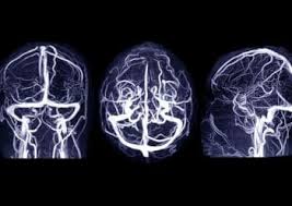
Medintu has collaborated with the best pathology laboratories that are NABL and NABH certified and follow ISO safety guidelines to provide the best MR Venography Brain at an affordable price for needy individuals. Magnetic Resonance Venography (MRV) of the brain is a unique imaging modality which offers detailed, noninvasive images of the brain veins. Unlike conventional MRI, MRS examines both the brain tissues and the blood vessel interior but MRV is confined only to the veins and venous sinuses in the brain. Since this technique employs Doppler signal analysis, it is most helpful in diagnosing conditions with respect to venous circulation, including cerebral venous thrombosis, venous malformations, dural arteriovenous fistulas and cerebral venous insufficiency.
MRV employs the most recent technology in MRI and may require that a fluid contrast agent be injected into the bloodstream to assist differentiate between normal and diseased veins in the brain. It has significant importance in assessment and treatment of those neurological disorders associated with venous blood flow and holds potential to replace the invasive diagnostic techniques such as catheter based venography. To schedule an appointment for MR Venography Brain, simply contact Medintu or call our customer care at +919100907036 or +919100907622 for more details and queries.
Overview
Specification
FAQ
Overview
Indications for MRI Venography of the Brain
MRI Venography of the brain is mostly useful in the diagnosis and assessment of diseases that involve veins in the brain. Below are the key indications for performing MRV:
- Cerebral Venous Thrombosis
Description: MRV can be used in diagnosis of such issues as cerebral venous thrombosis where blood clots are formed in the veins of the brain. It can lead to life threatening conditions including stroke, seizures, and raised intracranial pressure.
Why MRV: MRV also provides a great view of the venous sinuses so assessment of the blockages or clot that hinder the blood flow can be done properly.
- Venous Malformations
Description: MRV can be used to evaluate the venous malformation including Arteriovenous malformations or Dural arteriovenous fistulas.
Why MRV: It can help to define the size, location and extent of these vascular anomalies, and its mapping may be useful in planning treatment.
- Pulmonary Hypertension in Chronic Cerebral Venous Insufficiency
Description: MRI is employed to identify chronic cerebral venous insufficiency – a condition wherein blood flow from the brain is a problem, therefore resulting into complications including headache, dizziness and changes in cognition.
Why MRV: MRV may add visibility of the venous drainage pathways and their ability to help detect areas of blockage or constriction that may be hampering blood circulation.
- Stroke Investigation
Description: In stroke cases, MRV is used to find out whether it is due to venous pathology such as venous thrombosis or other angiopathy which led to reduced blood flow or ischemia.
Why MRV: The MRV also plays a role in excluding venous causes of stroke, including cerebral venous thrombosis, or in evaluating any venous factor that may contribute to the event.
- Possible Increased Intracranial Pressure
Description: MRV is useful in diagnosing intracranial hypertension which is an increased pressure of the brain.
Why MRV: An MRI can help determine whether the underlying problem is venous compression or other vascular disorders which may be leading to the high pressure in the cranial cavity.
How MRI Venography Works?
MRI Venography, or MRV of the brain, is a sophisticated scanning technique that uses magnetic fields and radio waves to visualize the venous structures inside the brain in a very detailed manner. Here’s how the procedure takes place:
- Basic MRI Principles
Magnetic Field: Similar to the convention MRI, MRV makes use of a very strong magnetic field that causes alignment in hydrogen atoms in the body. Human body, full of water, contains much hydrogen atoms; thus, perfect for MRI imaging.
Radio Waves: The radio waves travel in the body once the hydrogen atoms are aligned by the magnetic field. Then, the signals that come out from the hydrogen atoms are received by the MRI machine to create images of inner body parts.
MR Venography differs from a typical MRI in that it visualizes only the venous system of the brain rather than images of blood vessels and other tissues.
- Contrast Agent Administration
Contrast Injections: A contrast agent, usually a gadolinium-based contrast agent, is injected into a vein of the arm for enhanced visibility of the blood vessels, especially the veins. The contrast agent highlights the venous structures, making it easier to see the flow of blood and any abnormalities.
How It Works: Gadolinium Contrast Agent: Gadolinium makes the veins slightly less magnetic so that they stand out on an MRI as a brighter image; this enables the radiologists to separate them from brain tissues and arteries.
- Techniques of MRI Venography
There are two forms of MR Venography techniques :
Time-of-Flight (TOF) MRV: This technique is based on physiological motion of blood. This technique exploits the difference in behavior between flowing blood and stationary tissues and separates it from them by its velocity. TOF MRV is mainly used when a noncontrast study is desired.
Contrast-enhanced MRV: In the absence of detailed images, contrast enhancement on MRV is used. Via this technique, a contrast agent is administered to improve quality of images in venous structure. It is usually employed as a method in diagnosing abnormalities such as blood clot formation or venous malformation.
- Imaging Process
Place the patient on an MRI table and make his head available inside the machine. Sometimes a head coil is also provided with a head for high-quality images.
Scan Time: The MRI machine will produce a sequence of images of the venous system of the brain. Scanning time may take from 30 to 60 minutes, depending on the number of images needed. The patient must be motionless during the scanning process for clear and precise images.
Real-Time Visualization: The MRI machine captures multiple images of the brain during the scan, and a computer processes these images to produce high-resolution 3D images of the venous structures of the brain.
- Image Analysis
Once the images have been produced by MRV, a radiologist analyzes them to evaluate the anatomy of the veins and check for abnormalities such as thrombosis (blockage), AVMs (malformations), or narrowing of the veins.
MRV imaging allows for a three-dimensional reconstruction of the venous system, hence providing an overall view of veins and blood flow and anomalies of venous circulation in the brain.
Benefits of MRI Venography of the Brain
MRI Venography or MRV of the brain gives ample advantages to the patient as well as to the healthcare provider. Being known to have aided diagnoses for vascular conditions which predominantly occur within the venous system that do not entail an invasive procedure, some of the important advantages are the following.
- Not Invasive and thus, very safe.
Non-Invasive: Unlike classical venography, which employs a catheter passed into blood vessels, MRV does not require any surgical procedures and incisions.
No Ionizing Radiation: MRV will not use ionizing radiation as compared to CT scans or X-rays. This makes the procedure safer, especially if repeated imaging studies are expected or if the patient is pregnant.
- High-resolution imaging of venous structures
MRV also gives clear images of the veins and venous sinuses in the brain. One can even view a small anomaly like venous thrombosis, venous malformations, or DAVFs.
Full view: Imaging also gives full view of venous anatomy so that someone may have an image of the flow of blood inside the brain and know about those areas which have any obstacle or abnormality.
- Precise Vascular Conditions Diagnosis
Cerebral Venous Thrombosis: MRV is very useful in the diagnosis of cerebral venous thrombosis, a clot in the veins of the brain. Early detection can be lifesaving in preventing stroke and other serious complications.
Venous Malformations: AVMs or dural arteriovenous fistulas can be nicely visualized, and the physicians can judge the severity much better to develop an appropriate treatment strategy.
Cerebral Venous Insufficiency: An MRV can be useful to diagnose cerebral venous insufficiency, which is the failure of blood to drain out the brain. Symptoms can go from headaches, dizziness, up to impaired cognitive ability.
- No Invasive Tests Needed
No Invasive Catheterization: The process of traditional venography usually involves the catheterization of veins; however, it is risky and could lead to infections, haemorrhage, among other discomforts. MRV on the other hand has no form of invasive technique hence non-risky and pain free.
Few Complications : Being a non-invasive imaging, MRV has fewer complications such as catheter-induced infections among others and thus safer for patients
- Suitable in Monitoring the Progression of Treatment
Monitoring Vascular Malformations: MRV is valuable in the follow-up of a dural arteriovenous fistula or venous malformation to help practitioners monitor the changes and make modification of the treatment.
Monitoring Postsurgical Treatment: After brain vascular system intervention, such as surgery, the success of the treatment, appearance of complications, or confirmation that the veins are working well can be detected using MRV.
Common Conditions Diagnosed with MR Venography of the Brain
MRI Venography, or MRV, is a diagnostic tool that holds the strength to detect and assess various conditions that affect the venous structures in the brain. Some of the most common conditions diagnosed with MRV are as follows:
- Cerebral Venous Thrombosis (CVT)
Description: CVT is a blood clot formed in the brain veins leading to normal blood flow blockage. This condition may be very complicated leading to stroke and even damage to the brain.
Symptoms: CVT includes signs such as headache, facial swelling, seizures, and neurological deficiencies.
How MRV Assists: MRV is a brilliant technique that depicts clots in cerebral venous sinuses and veins with very nice images that assist in the sizing and localization of thrombosis.
- Venous Malformations (Vascular Malformations)
Definition: A malformation is a group of abnormal veins that form in such an abnormal fashion that blood cannot flow appropriately. Similar kinds of presentations can be experienced with conditions like AVMs or Cavernous Malformations.
Symptoms: These abnormalities lead to symptoms like headache, seizures, or even bleeding. Compression of the surrounding tissues may impair normal brain function.
How MRV Assists: MRV assists in diagnosing anomalous vein formation and provides a wide view of size and location of these anomalies, which might be of help in planning the treatment.
- Dural Arteriovenous Fistulas (DAVFs)
Description: DAVFs are abnormal connections between arteries and veins of the dura mater or the outer layer of the brain. Such fistulas tend to disrupt normal flow. They may lead to dangers such as hemorrhage in the patient.
Symptoms: Headache, pulsatile tinnitus, seizures, and neurological deficits.
How MRV Assists: MRV is very effective in the detection of DAVFs because it offers images with abnormal blood flow patterns characterizing these fistulas.
- Cerebral Venous Insufficiency (CVI)
Definition: CVI is a disease where the veins in the brain are not able to drain the blood properly, and thereby proper venous circulation cannot be established. This will lead to various neurological manifestations.
Headaches, dizziness, cognitive dysfunction, and sometimes, pressure in the head.
How MRV Is Helpful: It is helpful in the visualization of the blocked or narrowed veins and can identify the areas where the blood flow is not sufficient. This may lead to symptoms of CVI.
- Intracranial Hypertension or Elevated Intracranial Pressure
Definition: The intracranial pressure might rise due to different causes that include venous obstruction or malformation obstructing normal flow.
Common Symptoms: Major symptoms of such conditions would include the most severe form of headache, vomiting, abnormalities in vision, and altered consciousness.
How it would help with MRV: Maybe it might detect venous narrowing or obstruction, which in turn would cause increased intracranial pressure for the patient.
Specification
- Test Type: MR Venography Brain
- Preparation:
- Wear a loose-fitting cloth
- Fasting not required
- Carry Your ID Proof
- Prescription is mandatory for patients with a doctor’s sign, stamp, with DMC/HMC number; as per PC-PNDT Act
- Reports Time: With in 4-6 hours
- Test Price: Rs.8000
FAQ
How to book an appointment for a MR Venography Brain?
To schedule an appointment for MR Venography Brain, simply contact Medintu or call our customer care at +919100907036 or +919100907622 for more details and queries.
What is MRI Venography of the Brain?
This specialized form of imaging depicts the venous system of the brain in detailed images of veins and venous sinuses taken by magnetic fields and radio waves, thus enabling doctors to diagnose cerebral venous thrombosis, venous malformations, and other vascular abnormalities in the brain.
Why is MRI Venography done?
MRV is used mainly for diagnosing conditions of cerebral venous, including cerebral venous thrombosis, venous malformations, such as arteriovenous malformation or dural arteriovenous fistula, and other cerebral venous conditions. It is also used to diagnose chronic cerebral venous insufficiency and intracranial hypertension assessment.
What is the difference between MRI Venography and MRI?
Although a routine MRI gives a high-resolution view of the tissues and blood vessels in the brain, MRI Venography views the venous system. MRV uses special techniques, often with contrast agents, to look at veins and blood flow in detail and is mainly used for vascular diagnosis.
Is MRI Venography painful?
The MRI Venography is generally painless. However, the most uncomfortable thing is to lie there for a long time, usually 30-60 minutes. If a contrast agent is used, some may feel a mild warmth feeling when it is injected, but it usually isn’t painful.
Do I need to prepare for an MRI Venography?
Preparation for MRV is usually minimal. You would be asked to remove all your metal objects, for instance, jewelry since they would interfere with the MRI. If contrast is used, you are expected to tell your doctor if you have any allergies, particularly an allergy to contrast materials or iodine. At times, you are asked to avoid taking some food for a couple of hours before the test.
Is the contrast agent used in MRI Venography non-toxic?
Generally, gadolinium-based contrast agents used in MRI Venography are safe, although there is a small potential risk for patients who may have kidney problems or any allergic reaction. Your physician will review your medical record and determine if it would be safe for you.
Are there risks in doing MRI Venography?
MRI Venography is a noninvasive procedure with very minor risks. It does not expose the patient to any form of ionizing radiation like a CT scan or an X-ray, making it relatively safer than these options. However, in patients with allergies, it might occasionally cause a mild reaction in their bodies, or in sensitive kidneys, may lead to problems. Therefore, ensure to disclose any allergies or renal disease to your doctor prior to the scan.
How long does an MRI Venography take?
The procedure for MRI Venography generally takes between 30 to 60 minutes, although it depends on the complexity of the case and the amount of imaging needed. If extra sequences are required or if a few areas need to be imaged, it can take more than that.
Why Choose Medintu for MR Venography Brain?
Medintu is an online medical consultant, which offers home services not only in your city but also in all major cities of India, such as Hyderabad, Chennai, Mumbai, Kolkata and others. This makes it easy for us to work with diagnostic centers that boast of having the most accurate equipment. The customer service for booking the appointment of the services is available 24/7 and Medintu also comes with instructions. Medintu has not only the best diagnostic centres, but it offers them at very cheaper prices. If you have been tested, you can promptly schedule an appointment with a health care service through our list of skilled physicians. For appointment for MR Venography Brain, you can chat with us through Medintu or call our customer care at 919100907036 or 919100907622 for more information or inquiries.