


Fill out form to enquire now
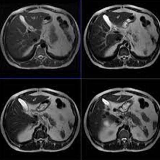
MRI Upper Abdomen and MRCP
Medintu has collaborated with the best pathology laboratories that are NABL and NABH certified and follow ISO safety guidelines to provide the best MRI MRI Upper Abdomen with MRCP at an affordable price for needy individuals. Upper Abdominal MRI scan is a useful tool in getting images of the internal structure of the upper abdomen which comprises the liver, the pancreas, the gallbladder and the biliary tract using Magnetic Resonance Cholangiopancreatography(MRCP). MRCP, a sort of MRI, has the special feature that directs it at the biliary and pancreatic duct and might be valuable in the visualization of pathology of these structures. This is essential in defining a number of diseases. MRI and MRCP offer one of the greatest benefits of being minimally invasive. There is little or no pain following these processes and no radioactive dangers anyway. This makes MRI particularly employable for repeat images and in pregnancy, children and any other susceptible group of people appropriately. When used in conjunction, MRI and MRCP provide a wealth of diagnostic data that allow for the proper diagnosis and treatment of some common abdominal disorders. This imaging method is not only useful in diagnostic purposes, but also useful in therapeutic intervention planning and also in the assessment of the success of on-going therapy.
To schedule an appointment for an MRI Upper Abdomen with MRCP, simply contact Medintu or call our customer care at +919100907036 or +919100907622 for more details and queries.
Overview
Specification
FAQ
Overview
What is an MRI?
The MRI is, therefore, a medical imaging technique that is used to make images of organs/tissues in the body. MRI is a technology that operates with strong magnets and radio waves in order to create images. According to this understanding, the MRT draws the hydrogen atoms in the body by dropping the magnetic field on them and through the radio waves interrupts this. And when the signal is switched off the hydrogen atoms release signals while getting back to normal position and these signals are used to build images. MRI uses high-resolution images that make it most suitable for capturing soft tissues, brain muscles and organs such as the thyroid gland. MRI is different from other techniques such as X-ray or computer tomography which utilise ionising radiation making MRI safer for many patients. MRI can be applied even to diagnose different disorders, such as tumours or flu, inflammation, and injuries. MRI is used to detect cancer, brain tumours, joint diseases, and spinal diseases. It can also be used to track the results of a disease or a treatment program.
What is MRCP?
MRCP is a MRI based imaging technique to obtain the visuals of biliary and pancreatic duct in a non-interventional manner. It is particularly useful for conditions involving liver and gallbladder, biliary tract and pancreas. MRCP is not an invasive procedure, and no surgical action is performed on patients, making it safer than any other procedure. This technique enables the capturing of high-contrast images in the biliary and pancreatic ductal systems used in diagnosis of diseases. A critical aspect of MRCP is it does not use ionizing radiation, unlike other related techniques like the computed tomography scans. MRCP can be used to diagnose bile duct obstruction, pancreatitis and its complications, congenital diseases of the biliary tract, stenosis or pathologies of the bile ducts and the pancreatic duct. MRCP is safely used in diagnosing diseases since it can detect any abnormalities in the biliary and pancreatic ducts at a very high sensitivity.
Indications for MRI of the Upper Abdomen with MRCP
MRI combined with MRCP is used in evaluating numerous diseases and pathologies located in the upper abdomen –like liver, pancreas, biliary system, etc. Here are some common indications for this combined imaging approach:
1.Biliary Obstruction:
To investigate the causes of obstruction of the bile ducts including gallstones, tumors or stricture.
2.Pancreatitis:
For the diagnosis of acute pancreatitis and chronic pancreatitis and also for the diagnosis of complications such as pancreatic necrosis or pseudocyst formation.
3.Tumors:
For the diagnosis and staging of tumours which may be primary or metastatic in the liver, pancreas or the biliary tree.
4.Cholangiocarcinoma:
When diagnosing cholangiocarcinoma and assign it into stages and when assessing the resectability of the tumour.
5.Congenital Anomalies:
In assessment of congenital abnormalities of the biliary or pancreatic ducts.
6.Infection or Inflammation:
In order to evaluate diseases like cholangitis, that is inflammation of the bile ducts, or abscess formation in the vicinity of the liver or pancreas.
7.Post-Surgical Evaluation:
If you are reading this page for advice on post-operative imaging after biliary or pancreatic surgery to look for complications including leaks or strictures, then you are in the right place.
8.Liver Disease:
For assessment of chronic liver pathologies such as cirrhosis, hepatitis etc, and their effects on biliary system.
9.Assessment of Vascular Structures:
To map relative positions and orientations of the hepatic artery, portal vein, and other vessels of the abdominal cavity in connection with abdominal organs.
10.Evaluation of Abdominal Pain:
Patients with chronic pancreatitis, suspected duodenal or periampullary malignancy, and those with normal pancreatitis enzymes.
11.Guidance for Interventions:
MRCP can be useful for planning intervention such as ERCP or biopsies, because MRCP displays the anatomical layout of the pancreas in exquisite detail.
Preparing for MRI and MRCP
MRI of the upper abdomen with MRCP requires proper preparation to avoid erroneous conclusions as well as patient discomfort. Here’s what you need to know:
- Dietary Restrictions:
Fasting: The colonoscopy may be performed after fasting for 4-6 hours especially if a contrast agent is to be administered. Consult your doctor to determine the proper way for you to take it.
Hydration: One normally can consume clear fluids until a few hours to the exam, depending on the doctor’s advice.
- Medications:
Continue Medications: Generally, all drugs can be used without restrictions, but always tell your doctor what drugs you are taking, including anticoagulants or drugs that affect kidney function.
Contrast Agent Considerations: Before the examination, it is necessary to tell your doctor if you have ever had any allergies to contrast agents or kidney problems.
- Clothing:
Dress Comfortably: It is advised patients wear comfortable, baggy garments which do not contain metal clasps (zippers, buttons) for the procedure. Occasionally, you might be required to wear the hospital garment known as a gown.
- Metal Objects:
Remove Metal Items: In preparation for the MRI, there are several accessories and clothing that will need to be left outside the theater; belts, watch, jewelry, hairsticks, and metal clothing. This is important because the use of metal may confound the magnetic field and imaging.
- Health History:
Inform Your Provider: Some questions include; Do you have any other medical conditions? Any kidney related issues, liver issues or any previous abdominal surgery? Also it is recommended to tell the doctor if you are pregnant or if you may be pregnant.
- Claustrophobia and Anxiety:
Address Concerns: If for instance you are a claustrophobia, that is, if you fear tight spaces, you should tell your healthcare provider. Possibly, they will suggest something to ease your discomfort, including an administration of some sedatives or the use of calming breathing patterns.
- Arriving for Your Appointment:
Plan Ahead: Get to the imaging facility about 15- 30 minutes earlier to fill out any forms that you might have to fill if needed for the scan.
The MRI and MRCP Procedure
- Arrival and Check-In:
There may be tradition to come to the imaging facility 15-30 min. before the appointment to complete the forms and to prepare for the examination.
- Preparation:
Changing Clothes: Sometimes you may be asked to change into a hospital gown so that nothing you wear has metal on it.
Removal of Metal Items: Ensure that all objects that are nice or look new such as rings, necklaces, belts or anything metallic is removed before going for the MRI scan.
- Getting Comfortable:
Positioning: The test is performed when the patient is lying on the bed with a top layer of a cushioned examination table. Sometimes it may happen that because of the part of the body to be imaged, you may be asked to lie on your back or on your side.
Safety Checks: Through such assurances one may safely conclude that a technician gives a medical history check and then makes sure that he or she is alright in the position in which he or she is receiving the procedure.
- The MRI Scan:
Entering the MRI Machine: Standard operating table will be used, and will be on wheel to transfer it inside MRI machine which is a huge cylindrical magnet. It may sound like an inconvenience, but there will be the ability to constantly talk to the technician during the procedure.
Imaging Process: The MRI will take images of your upper abdomen as well with a lot of detail. During the scan, you will hear loud tapping or thumping sounds, which is quite alright. Earplugs or headphones might be made available to minimise the disturbance.
- MRCP-Specific Imaging:
Specialized Sequences: For the MRI we will add sections to cover the bile ducts and the main pancreatic duct. This can entail other sequences that target these structures by themselves.
Breath-Holding: Sometimes you are required to close the mouth and take short breaths during some images to avoid blury images.
- Completion of the Procedure:
Duration: From start to finish the MRI and MRCP can take anywhere from 30 to 60 minutes depending on the specifics of the scans.
Post-Procedure: Then of course in the morning after the scan is done you can sit up and even get dressed. If a contrast agent was given you will be advised to drink fluids to eliminate it from your body.
Interpreting MRI and MRCP Results
- Image Acquisition:
MRI and MRCP results are the sets of images in axial, sagittal, coronal planes that give the clear depiction of the upper abdominal organs and structures.
- Role of the Radiologist:
Specialized Training: An MRI scan and an MRCP scan are combined and the images are analyzed by a radiologist, a physician specializing in interpreting pictures of the human body. This way they look for any structural and/or functional alteration of the organs within the chest.
Comparative Analysis: The radiologist may even look at the present images with those of previous imaging studies to determine changes.
- Key Factors Analyzed:
Biliary Ducts: The radiologist evaluates the biliary system for evidence of blockage, narrowing, or pathology that includes gallstones, tumor, or cysts.
Pancreatic Ducts: Assessment of the blood supply of the pancreas, angiogram to detect infiltration and signs of ischemia, require for such conditions as pancreatitis or tumors.
Surrounding Structures: Scanning of other organs which are close to the colon ; liver and gallbladder and kidney to check for any complications or associated diseases.
- Common Findings:
Normal Results: In the case where no pathology is seen and there is a negative MRI and MRCP the report will clearly state this as normal anatomy and function.
Abnormalities Identified:
Obstructions: Stones in the system include stones in the bile ducts, or stones in the pancreatic ducts.
Tumors: Identification of masses in liver, pancreas or bile duct, description of the mass and its spread.
Inflammation: Symptoms of pancreatitis, shown by inflammation or fluid accumulation, cholangitis.
Anomalies: Anomalies of the biliary system that may be present at the moment of birth or soon after.
- Reporting the Results:
Radiology Report: The radiologist summarises the concerned findings directly into a written report which may contain a diagnosis if there is one as well as the impression and recommendations for extra investigations or management in case of need.
Risks and Considerations
- Magnetic Field and Metal Objects:
Implanted Devices: Men with a penile implant or a man with certain metal ordinally in the body such as pacemaker or other metal objects such as clips or metal stents in the body cannot undergo MRI examination.
Screening Process: The patient fills a screening questionnaire that helps the doctor to determine whether the patient is fit for the procedure or not.
- Contrast Agent Reactions:
Use of Contrast Agents: Using a contrast agent if images have to be enhanced can be risky as it may cause allergic reactions. For the most part it only causes minor symptoms (itching, rash) in the majority of patients but in a very small percentage of cases it may cause life threatening symptoms (anaphylaxis).
Nephrotoxicity: Patients with history of renal disease should be screened because some contrast agents are associated with NSF.
- Claustrophobia and Anxiety:
Closed Space: Some of the patients might develop some form of anxiety and claustrophobia when they are inside the MRI equipment. Concerning the possibilities of using sedatives or techniques that can help to relax during a difficult period, it is possible to consult the doctor beforehand.
- Pregnancy:
Pregnancy Considerations: MRI is not used in pregnancy unless necessary and it is rarely used during the first trimester despite its safety during pregnancy. It is always important to report any pregnancy or possible pregnancy to your health care provider.
- Procedure Discomfort:
Positioning: Staying in a single position for a relatively long time might be disastrous to the body and can be very painful to the affected person, depending on his or her health complications. Telling the technician about it can help the patient receive adjustments or breaks in the course of the procedure.
- Radiation Exposure:
No Ionizing Radiation: MRI and MRCP are safety imaging techniques since they do not emit ionizing radiation as in the CT scans. However, the overall radiation risk should be remembered if the patient may have had a previous imaging exposure.
- Post-Procedure Monitoring:
Hydration After Contrast: If a contrast agent is used, then drinking much water after the procedure will help to clear it from the organism and prevent kidney problems.
Specification
- Test Type: MRI Upper Abdomen with MRCP
- Preparation:
- Wear a loose-fitting cloth
- you might need to stop eating and drinking around 4 to 5 hours beforehand
- Carry Your ID Proof
- Prescription is mandatory for patients with a doctor’s sign, stamp, with DMC/HMC number; as per PC-PNDT Act
- Reports Time: With in 3-4 hours
- Test Price: Rs.5500
FAQ
How can I book an appointment for an MRI Upper Abdomen with MRCP through Medintu?
To schedule an appointment for an MRI Upper Abdomen with MRCP, simply contact Medintu or call our customer care at +919100907036 or +919100907622 for more details and queries.
Can anyone give me a brief on what MRI is and also what is the difference between MRI and MRCP?
MRI is a common technique in diagnosing diseases in different parts of the body while MRCP is a specific MRI that studies the biliary and pancreatic ducts.
What is the duration of the said process?
Both MRI and MRCP usually require about 30 to – 60 minutes. The exact time therefore depends on the scan that has been done as well as the particular sequence that was employed.
Are MRI and MRCP procedures painful?
More importantly, the procedure is ordinarily not uncomfortable in any way. But, like in any procedure some patients may feel uncomfortable from the position they adopt during the procedure or from the injection of a contrast solution.
Is it necessary to avoid food before the procedure is administered?
Yes, you may be asked to appease for 4-6 hours prior to the MRI/MRCP halves if a contrast agency is to be administered. In any case, the instructions that you have to follow should be clear and specific at the same time given by your doctor.
What should I wear to the appointment..?
Avoid the use of clothes that have parts made of metals. You might be required to undress for the procedure, and wear a hospital gown in their place.
What are the dangers of MRI and MRCP?
MRI and MRCP are usually safe methods, but they have contraindications having to do with implanted devices, contrast agents, and having claustrophobia. Any other issues could be discussed with your doctor before the procedure.
What happens if I have a contrast allergy?
If you have a known allergy to contrast agents inform your doctor. They may have to employ other imaging techniques or give you some medication before the test to reduce the possibility of a reaction.
Is it safe to undergo an MRI if I am pregnant?
MRI is contraindicated in the first trimester unless the benefits of performing the study outweigh the risks of exposure to radiation. It’s important you always tell your doctor if you are pregnant or if you think there’s even a slight chance you could be.
Why Choose Medintu for MRI Upper Abdomen and MRCP?
Medintu is an online medical consultant that provides home-based medical services not only in your area but also in most cities in India, including Hyderabad, Chennai, Mumbai, Kolkata, and more. We have collaborated with diagnostic centers that have the best machines and equipment to ensure you get accurate results. Medintu provides 24-hour customer service for booking the appointment of the services and guides you with instructions. Medintu also provides the best diagnostic centers at low prices. Once you receive your test results, you can easily book an appointment with our network of experienced doctors for consultation. To schedule an appointment for an MRI Upper Abdomen with MRCP, simply contact Medintu or call our customer care at +919100907036 or +919100907622 for more details and queries.