


Fill out form to enquire now
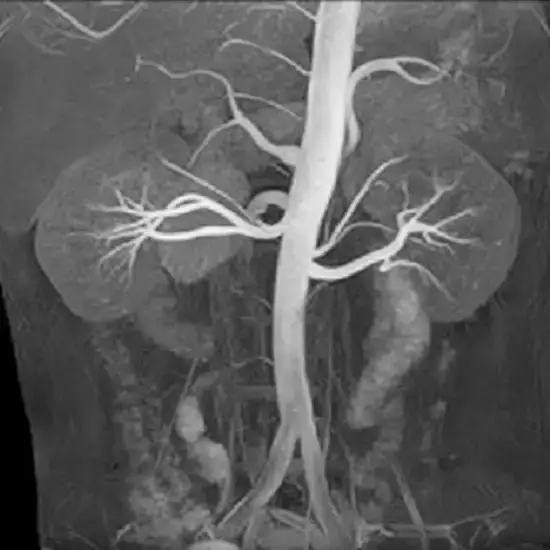
MR Renal Venography
Our advanced MR Renal Venography services at Medintu provide accurate imaging of the surrounding blood arteries and renal veins. Strong magnetic fields and radio waves are used in this non-invasive treatment to diagnose disorders such as varicocele, vascular compression syndromes, and renal vein thrombosis without surgery. Physicians will identify intravascular clots, diagnose conditions like nutcracker syndrome, and schedule preparatory treatments for kidney operations using our high-resolution imaging. Our MR Renal Venography technique ensures safer imaging, especially for patients requiring repeated scans, as it does not involve radiation exposure, unlike other methods such as CT renal Venography. At Medintu, we put patient safety first. For more information about MR Renal Venography cost visit Medintu or call us at +919618573143, +919100907622.
Overview
Specification
FAQ
Overview
What is MR Renal Venography?
MR Renal Venography (Magnetic Resonance Renal Venography) is magnetic resonance imaging of the renal veins and other vessels within or adjacent to the kidneys. It is helpful, especially in the conditions associated with disturbance of blood flow within the kidneys. This is a useful technique for assessing blood clots, venous malformations, and vascular compression syndromes involving the kidneys without needing surgery.
Why is MR Renal Venography Performed?
Conditions Diagnosed with MR Renal Venography: It is especially useful in evaluating pathological conditions affecting the veins with special emphasis on renal vein thrombosis syndrome, compressive syndromes, and varicocele.
Evaluating Renal Vein Thrombosis: MR Renal Venography enables the identification of intravascular thrombus formation and this can pose a risk to renal function due to thromboembolic-sudden renal failure.
Understanding Renal Vein Compression Syndromes: Nutcracker syndrome wherein the left renal vein is situated between the superior mesenteric artery and the aorta is one of the examples that can be revealed with this imaging technique.
Preoperative Planning for Kidney Surgeries: To determine the vascular structure surrounding the kidneys, surgeons use MR Renal Venography, which is essential in organizing kidney transplant operations.Understanding MR Renal Venography
Overview of the Imaging Process: MR Renal venography is imaging of the renal veins using strong magnetic fields, radio waves and a computer. The patient undergoing the scans is placed within the spherical MRI scanner and images are captured.
Use of Contrast Agents in MR Renal Venography: A gadolinium-based contrast material is also administered to the subject to enhance the vessels including the renal veins.
Key Features of Magnetic Resonance Imaging: Different from X- rays and CT scans, MRI discards the use of penetrating radiation as MRI uses magnetic and radio frequency technology. It takes high definition images and is hence suitable for kidneys where malformations, soft tissues and blood vessels should be visualized.Benefits of MR Renal Venography
High Resolution Imaging: The MR Venography verbalizing renal veins obtains a clear imaging of internal structures allowing precise problem identification of the veins.
Non invasive and Safe: This is a non invasive procedure that involves no invasion of the body and thus no surgical cuts are required. It is safe for most patients’ subjects and hence no complications are usually noted.
No Radiation Exposure: Since MR Renal Venography utilizes magnetic fields rather than X-rays, there is no exposure to ionizing radiation hence there are safety levels, particularly for patients requiring serial imaging.Preparing for an MR Renal Venography Scan
Pre-Procedure Guidelines: Patients can be instructed not to eat or drink for a couple of hours before the procedure in a case where contrast will be injected. It is also necessary to inform the physician of any present disease or allergy of the patient.
What to Expect During the Procedure: Hospitals are filled with mental images of patients being wheeled into surgery wearing only a hospital gown. During the scan, the patient remains in the MRI machine whereby the machine captures images of the patient’s renal veins. Usually, this procedure lasts 30-60 minutes. If contrast is used, it will be injected during the scan intravenously.Understanding the Results of MR Renal Venography
Interpretation of the Imaging Results: Images obtained after the completion of imaging are then evaluated by the radiologist for the presence of any disease in the renal vein. These results can be associated with the presence of such conditions as thrombosis, compression, and abnormal venous anatomy.
Common Findings and Their Significance: Abnormalities may be found on imaging, for example, renal vein thrombosis, Sugeryigan syndrome, or other vascular dysplasias. Based on these findings, the clinician can formulate a treatment plan for the patient.Potential Risks and Considerations
Possible Side Effects of Contrast Agents: Gadolinium-based contrast agents are highly tolerated by patients, however, some may experience mild reactions such as nausea or headaches. Allergic reactions can extremely infrequently take place.
Who Should Avoid MR Renal Venography?: Patients with specific implantable prostheses, namely, pacemakers, cochlear implants, and other devices, cannot perform the procedure since there is a strong magnetic field during the procedure for imaging. Pregnant women would also require a consultation with their physician before taking the scan.Comparison with CT Renal Venography
CT Renal Venography is another method of renal imaging that involves the use of computed tomography (CT) and contrast dye to visualize the renal veins. However, it involves radiation exposure, which is why certain patients will prefer MR Renal Venography.
Other Imaging Options for Renal Veins
The renal veins may be studied by ultrasound and Doppler studies, though these may be inadequate when compared with MR Renal Venography. Imaging method selection varies with the patient’s status and how detailed the images one seeks.
Specification
- Test Type: MR Renal Venography
- Preparation:
- Wear a loose-fitting cloth
- Fasting required
- Carry Your ID Proof
- Prescription is mandatory for patients with a doctor’s sign, stamp, with DMC/HMC number; as per PC-PNDT Act
- Reports Time: With in 4-6 hours
- Test Price: Rs.8000
FAQ
What is MR Renal Venography?
MR Renal Venography is a magnetic resonance imaging (MRI) technique that creates images of the renal veins and surrounding blood vessels. It is used to identify conditions like blood clots, venous malformations, and vascular compression syndromes affecting the kidneys.
Why would a doctor recommend MR Renal Venography?
Doctors may recommend MR Renal Venography to diagnose conditions like renal vein thrombosis, nutcracker syndrome, and other vascular abnormalities near the kidneys. It helps in evaluating blood flow disturbances and is crucial in preoperative planning for kidney surgeries.
How does MR Renal Venography work?
This technique uses strong magnetic fields and radio waves to capture high-definition images of the renal veins. Sometimes, a gadolinium-based contrast agent is injected to enhance the visibility of the veins during the scan.
Is MR Renal Venography safe?
Yes, MR Renal Venography is generally safe. It is non-invasive and does not use ionizing radiation like X-rays or CT scans, making it suitable for patients who need repeated imaging.
Are there any risks associated with MR Renal Venography?
Rarely, some may experience mild reactions like nausea or headaches due to the contrast agent. Severe allergic reactions are extremely rare. Always inform your doctor of any known allergies.
How do I prepare for an MR Renal Venography scan?
Your doctor may ask you to avoid eating or drinking for a few hours before the scan, especially if a contrast agent is used. Inform your doctor about any existing medical conditions or allergies before the procedure.
Can pregnant women undergo MR Renal Venography?
Pregnant women should consult their physician before undergoing MR Renal Venography. While MRI is generally considered safe during pregnancy, the use of contrast agents might require special consideration.
How does MR Renal Venography compare to CT Renal Venography?
MR Renal Venography is different from CT Renal Venography as it does not use radiation. This makes it safer for patients who need multiple scans. CT Renal Venography, however, can be quicker but involves exposure to ionizing radiation.
How long does it take to get the results of an MR Renal Venography?
After the scan, a radiologist will interpret the images, and your doctor will review the findings with you. The time to receive results can vary but results will be available within a few days.
Who analyzes the results of MR Renal Venography?
A radiologist interprets the images from MR Renal Venography and looks for any abnormalities. Based on these results, a nephrologist, urologist, or vascular surgeon may be involved in your care plan.
Where can I get MR Renal Venography done?
At Medintu, we offer advanced MR Renal Venography services. For more information on the procedure and cost, you can visit our website or contact us directly at +919618573143, or +919100907622.
Why Choose Medintu for MR Renal Venography?
Medintu helps you get the best and most accurate MR Renal Venography. We work with experienced radiologists and the best labs with top-quality MRI equipment. All our labs are certified by NABL and follow ISO guidelines so that we give you top-quality health care. Accurate diagnosis is ensured by our doctors at convenient locations of our centers not only in your area but also in most cities in India, including Hyderabad, Chennai, Mumbai, Kolkata, and more. Medintu provides 24-hour customer service for booking the appointment of the services and guides you with instructions.